|
PRACTICAL
SIGNIFICANCE
This
study
revealed
that
differences
exist
between
sexes in
spinal
control
of
movement
as well
as the
activation
of the
neuromuscular
system.
This
information
can be
used to
develop
interventions
and
training
programs.
STUDY
BACKGROUND
Females’
display
different
lower
extremity
movement
patterns,
particularly
at the
hips and
knees,
when
compared
to
males.
These
movement
patterns
potentially
put
females
at
increased
risk for
lower
extremity
injury.
However,
the
underlying
neural
mechanisms
controlling
these
movement
patterns
are
unknown.
Specifically,
differences
in motor
neuron
excitability,
intrinsic
and
extrinsic
pre-synaptic
inhibition,
postsynaptic
inhibition,
and
supraspinal
drive
between
the
sexes
are
unidentified.
Additionally,
sex
differences
in rate
of
torque
development
and
electro-mechanical
delay
are not
fully
elucidated.
OBJECTIVE
To
determine
if sex
differences
exist
between
spinal
control
mechanisms
and
functional
neuromuscular
variables.
DESIGN
AND
SETTING
A
between-subjects
design
was used
to
compare
differences
in rate
of
H-reflex
excitability,
rate of
intrinsic
pre-synaptic
inhibition
(IPI),
rate of
extrinsic
pre-synaptic
inhibition
(EPI),
level of
postsynaptic
recurrent
inhibition
(RI),
level of
supraspinal
neural
drive
(V-wave),
maximum
rate of
torque
development
(RTD),
and
electromechanical
delay (EMD)
between
healthy
active
males
and
females.
The
study
took
place in
a
controlled
laboratory
setting.
SUBJECTS
Nineteen
males
(age =
23.0 ±
4.3yrs,
height =
177.45 ±
5.44cm,
mass =
77.52 ±
13.18
kg) and
18
females
(age =
24.7 ±
2.9yrs,
height =
165.31 ±
5.85cm,
mass =
62.44 ±
8.76 kg)
healthy,
physically
active
individuals
participated
in the
study,
MEASUREMENTS
Participants
were
seated
on the
chair of
the
Biodex
System 3
dynamometer
in a
semi-recumbent
position
with the
knee
flexed
to 60
degrees
and
ankle in
anatomical
position.
All
subsequent
testing
was
performed
in this
position.
Hreflex
recruitment
curves
of the
soleus
were
elicited
at the
tibial
nerve.
IPI
recruitment
curves
were
obtained
by
utilizing
paired
pulses.
Two
stimuli
of the
same
intensity
and an
interstimulus
interval
of 100
ms were
given to
the
tibial
nerve.
To
obtain a
EPI
recruitment
curves,
the
H-reflex
was
conditioned
by
stimulating
the
tibialis
anterior
(50% TA
Mmax)
100 ms
prior to
stimulating
the
soleus
muscle.
The
first
derivative
of all
three
recruitment
curves
was
calculated.
RI was
determined
by the
difference
between
S1
alone,
which
was 25%
of
soleus
Mmax,
and S1
conditioned
by S2,
which
was set
to Mmax.
Ten
trials
of each
were
completed.
V-waves
were
elicited
by a
supramaximal
stimulation
to the
tibial
nerve
once 90%
of
maximum
isometric
plantarflexion
torque
was
reached.
Five
trials
with 60
seconds
rest
were
obtained.
To
measure
soleus
RTD and
EMD
participants
were
instructed
to
isometrically
plantarflex
the
ankle as
fast and
as hard
as they
could.
Three
trials
with 60
seconds
rest
were
measured.
EMD was
determined
by
calculating
the time
between
the
onset of
soleus
EMG
activity
and
onset of
soleus
torque
production.
RTD was
calculated
by
determining
the
slope of
the
torque-time
curve
from the
onset of
torque
production
to the
maximal
torque
production.
RESULTS
The
Wilks
Lambda
multivariate
test
revealed
differences
between
sexes on
the
linear
combination
of motor
neuron
pool and
functional
neuromuscular
variables
(P
=
0.001).
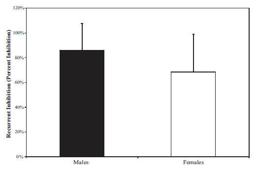
Follow-up
univariate
tests
revealed
that
males
had
significantly
greater
RI
(males =
0.86 ±
0.21,
females
= 0.68 ±
0.30;
P =
0.042;
See
Figure
1).
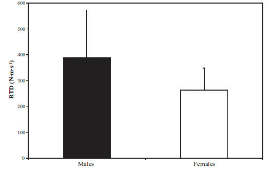
Males
also had
greater
RTD
(males =
387.93 ±
180.90
n·m·s-1,
females
= 263.89
± 85.15
n·m·s-1;
P=
0.033;
See
Figure
2).
No other
univariate
tests
were
significant.
CONCLUSIONS
The
sexes
differ
on
modulation
of
spinal
control
of
movement
and
activation
of the
neuromuscular
system.
Males
were
able to
produce
torque
more
quickly
than
females.
This is
potentially
important
during
injurious
situations
when
rapid
activation
of the
muscle
is
needed
due to
the
length
of time
needed
to
attain
maximal
torque.
Additionally,
RI was
greater
in
males.
RI is
considered
to be a
variable
gain
control
by
modulating
motor
unit
firing
frequency.
Both of
these
variables
are
modifiable
with
training.
However,
no
differences
were
observed
between
the
sexes on
motor
neuron
excitability
or
presynaptic
inhibition.
Figure
1:
Recurrent
inhibition
between
the
sexes
Males
demonstrated
significantly
more
recurrent
inhibition
compared
to
females
(P
=
0.042).
Figure
2:
Rate of
torque
development
between
the
sexes
Males
demonstrated
significantly
great
rate of
torque
development
compared
to
females
P=0.033.
|
Publication
&
Presentation
List:
-
Johnson S, Hoffman MA. Spinal Control Differences between the Sexes. NATA Annual Meeting and Clinical Symposium. San Antonio, Texas, June 17-20, 2009.
|
|
|
|

Sam Johnson, PhD, ATC
Principal Investigator
|
Sam Johnson, PhD, ATC
Sam Johnson recently completed his PhD at Oregon State University. He earned his Master’s in Kinesiology from the University of Nevada, Las Vegas and his Bachelor’s of Science from Texas Christian University. He has also worked as an Athletic Trainer at the University of Portland and Stanford University. |
|
|
This
Grant
Information
Summary
may be
downloaded
in a
2-page
pdf file
from
http://www.natafoundation.org/wp-content/uploads/2012/11/Johnson09.pdf |
Back to
December 19, 2012
eBlast
Newsletter
Send e-mail
to rachaelo@nata.org with questions
or
comments
about this web site. |