Manual Muscle Testing of the Cervical Spine
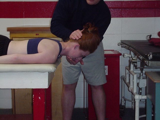
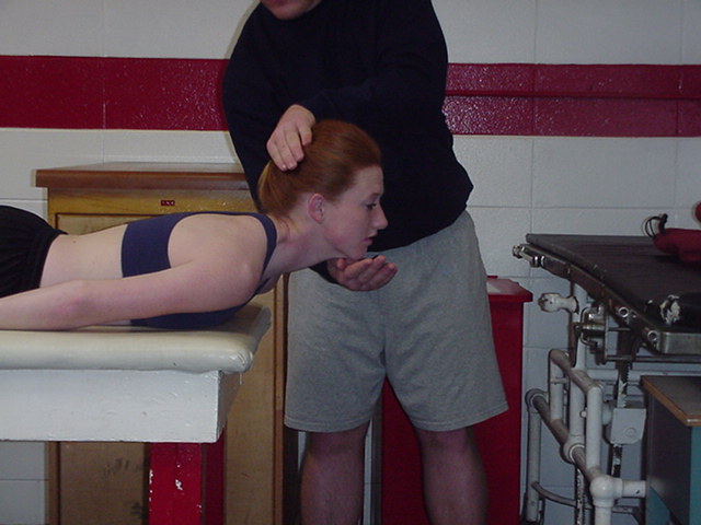
| Capital Extension | Model is prone with head off the end of the table. model's arms are at his side. The examiner stands at the side of the model next to the head. The examiner applies resistance with one hand over the occiput, and the other hand is placed under the head should it give way with applied resistance. | |
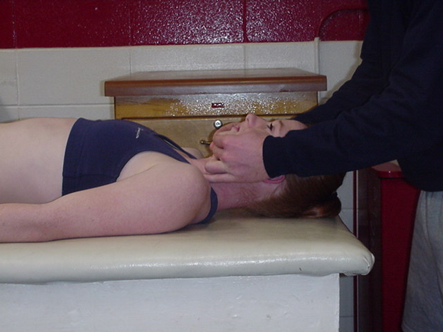
| Capital Flexion | Model is supine with head on table and arms at side. The examiner is standing at head of table facing patient. Both hands of examiner are cupped under the mandible (chin) and touching the cheeks to give resistance in an upward and backward direction. | |
| Cervical Extension | Model is prone with head off the end of table and arms at side. The examiner stands next to patients head. The examiner places one hand over the parieto-occipital for resistance and the other hand is placed under the chin ready to catch the head if it gives way. | |
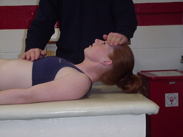
| Cervical Flexion | Model is supine with arms at their side, and head on table. The examiner stands next to patients head. The examiner places one hand on the model's forehead for resistance, and the other hand may be placed on model's chest. Stabilization is only needed when trunk is weak. | |
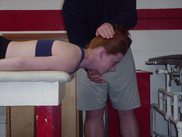
| Capital plus Cervical Extension | Model is prone with head off the table and arms at side. The examiner stands next to patients head and places one handover parieto-occipital area to apply downward and forward resistance. The other hand is placed under the chin to catch the head if it gives way. | |
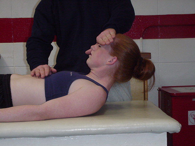
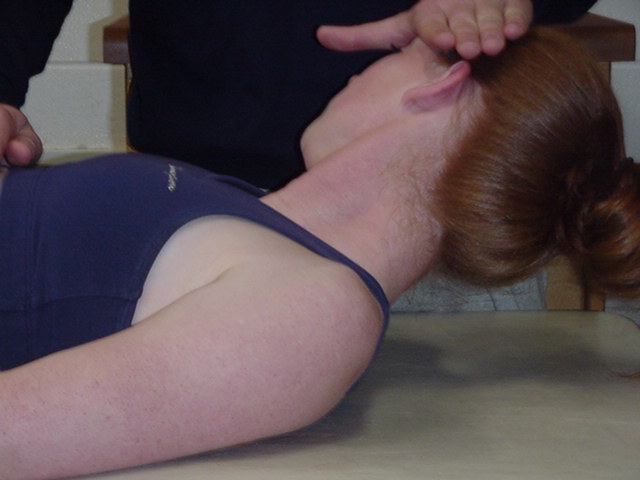
| Capital plus Cervical Flexion | Model is supine with head on table and arms at side. The examiner stands at the side of the table at the level of the model's shoulder. One hand is placed on the forehead for resistance and the other hand may be used for stabilization of the trunk if it is weak. | |
| Neck Flexion with Rotation | Patient is supine with head supported on table and turned to the right to test the left sternocleidomastoid. Therapist faces the patient with one hand placed on the temporal area above the ear for resistance. Patient raises head from table against resistance, keeping head turned throughout the movement. | |
Adapted from:
Hislop, Helen J. & Montgomery, Jaqueline with contributor Barbara Connelly.
Daniels and Worthingham's muscle testing: techniques of manual examination., 6th edition, 1995.