|
Bony Landmark
(include alternative name if applicable)
|
Related Information
such as purpose, function,
attachment of ligaments, tendon, soft tissues involved
|
Preferred Body &
Joint Position
best for palpation
|
Anatomical
Description of Location
relative to other structures
|
Skeleton Picture or Video
|
Model Picture or Video
|
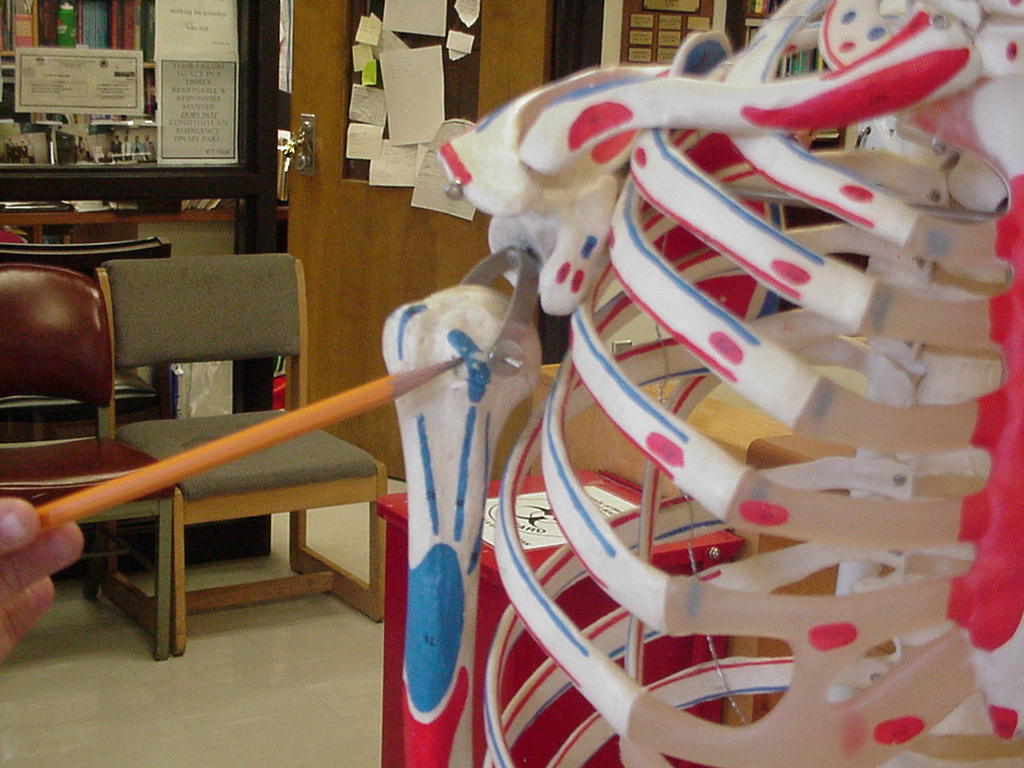
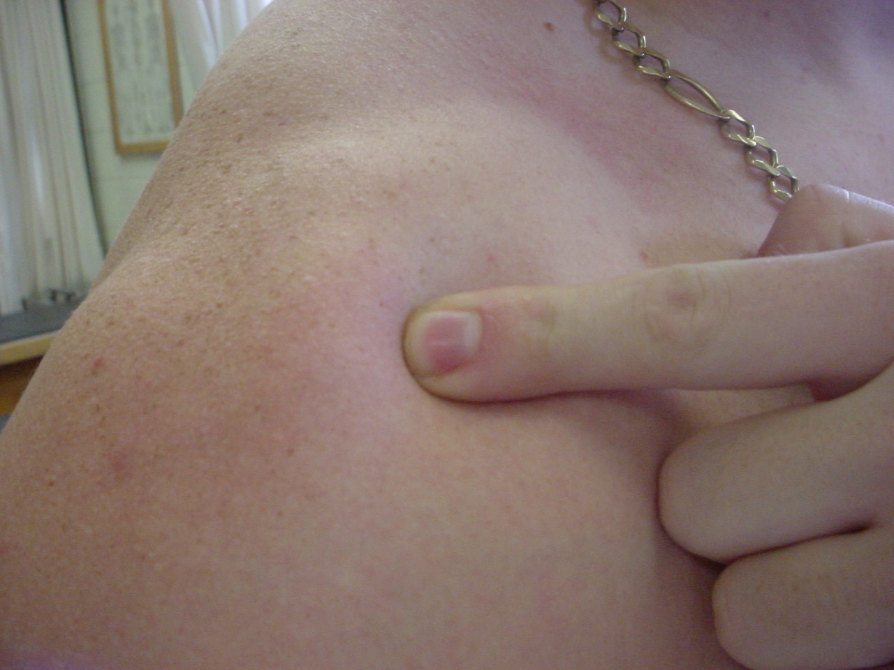
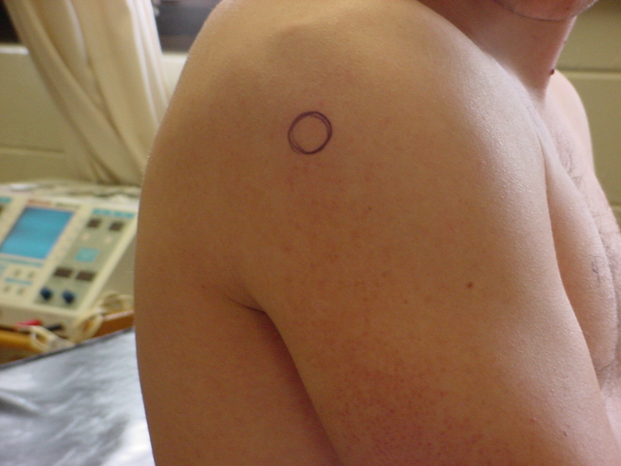
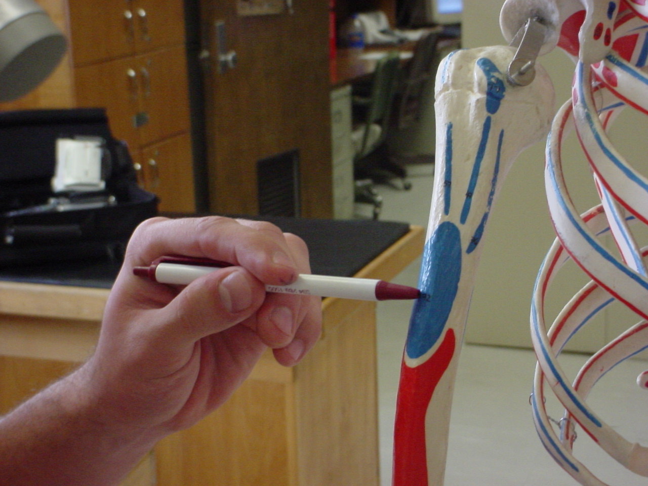
| Humeral greater tubercle |
Serves as an attachment for the anterior
circumflex humeral artery |
Patient sitting in a chair with the examiner
standing behind them |
Located laterally to the lateral tip of the
acromion |
 |
|
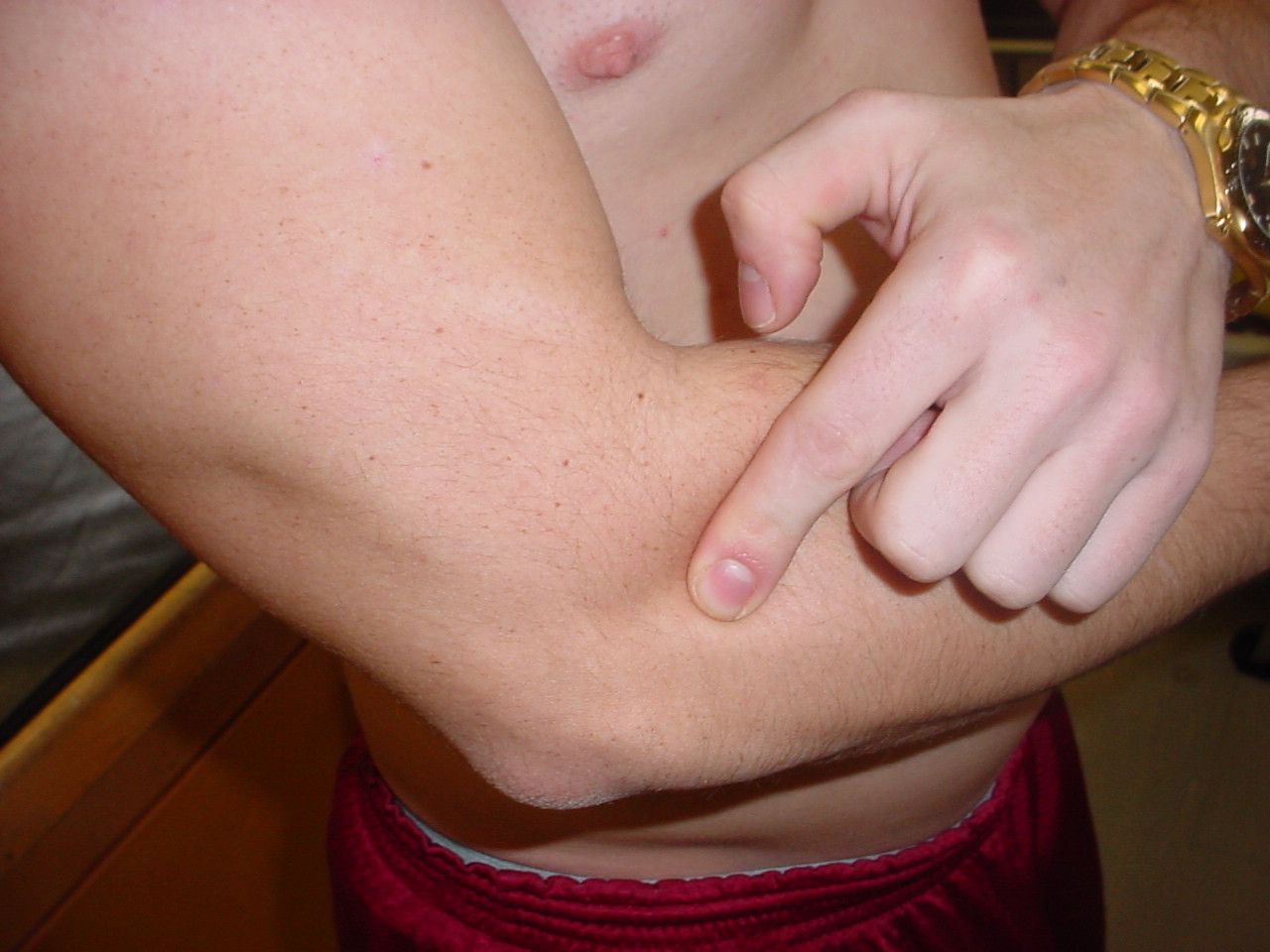
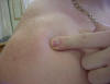
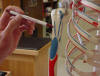
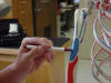
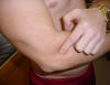
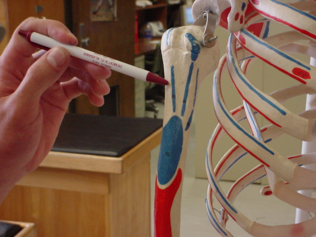
| Humeral lesser tubercle |
Is the attachment
for the subscapularis and to its sharp lateral margin the transverse
ligament |
Elbow flexed to 90
degrees; the examiner will place his/her finger into the bony prominence
of the bicipital groove; The examiner will then externally rotate the arm
and a bump will be felt, the examiners finger should feel another bump
that is the lesser tubercle; To better locate the lesser tubercle you can
internally and externally rotate the shoulder |
The lesser tubercle
is on the anterior aspect of the humerus bone imediately beyond the
anatomical neck, and shows a smooth, muscular impression on its upper part |
 |
_small.JPG) |
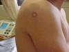
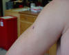
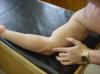
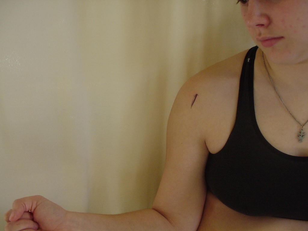
| Humeral head |
|
Sitting or supine
with arm relaxed |
Proximal end of the
humerus; fits into glenoid cavity of scapula |
_small.JPG) |
 |
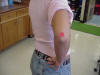
| Humeral neck |
Serves as an attachment for the lesser tubercle |
Patient should be standing with arm slightly
abducted and relaxed |
The neck is just inferior to the humeral
head |
 |
 |
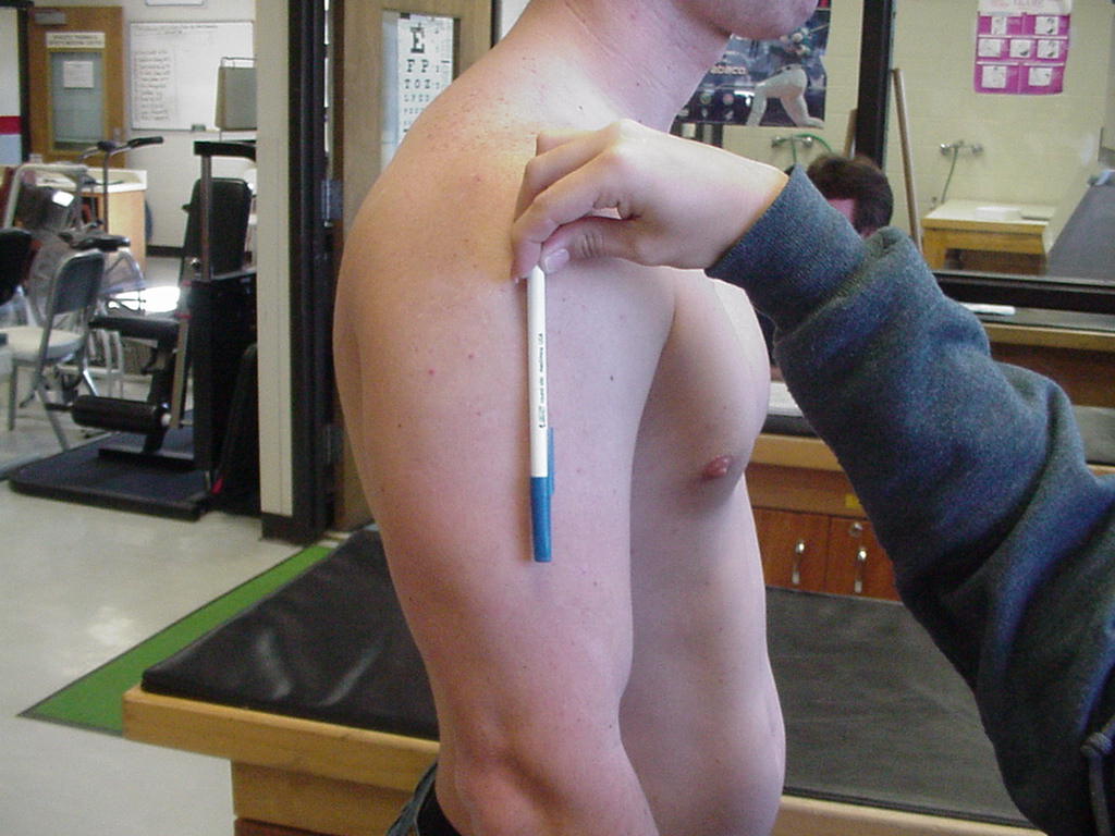
| Humeral shaft |
The humerus serves
as the insertion of the deltoid tuberosity |
The patient can be
standing or seated, facing the examiner with shoulders in neutral position |
Located distal to
the glenohumeral head and proximal to the medial and lateral epicondyles |
 |
 |
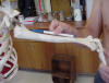
| Intertubercular groove,
bicipital groove |
Attachment site of
teres major, latissimus dorsi, and pectoralis major |
Arm in external
rotation, patient sitting or standing |
Bordered laterally
by the greater tuberosity and medially by the lesser tuberosity |
_small.JPG) |
 |
| Deltoid tuberosity |
Serves as the insertion for the deltoid muscle |
Patient standing and relaxed |
Located superior to the triceps muscle |
 |
 |
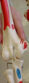
| Olecranon fossa |
It houses the
olecranon of the ulna; part of the triceps brachii tendon covers the
superior ridge of the fossa |
Place the patient
in anatomical position with the elbows flexed about 20 degrees |
Palpate just below
the posterior distal end of the humerus; This fossa is where the proximal
end of the ulna sits when the elbow is fully flexed |
 |
 |
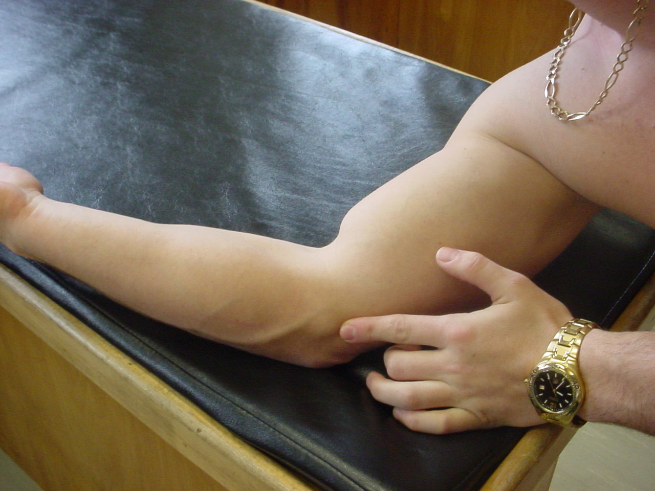
| Medial epicondyle |
Attachment site of
forearm flexors and forms the cubital tunnel |
Slight elbow
flexion |
Distal, medial end
of humerus |
 |
 |
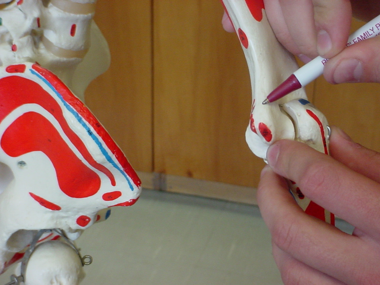
| Lateral epicondyle |
Serves as the site of attachment for the
lateral collateral ligament |
Patient standing, relaxed with arm flexed to 30
degrees |
Located lateral to the olecranon process |
XXX |
 |
| Cubital tunnel |
Triangular space
bordered laterally and medially by the pronator teres |
Elbow flexed at approximately 30 degrees |
Located between the two epicondyles of the
humerus |
|
|
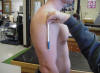
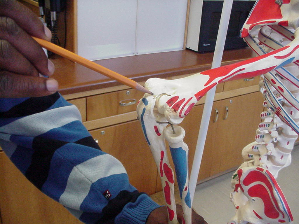
| Capitulum |
Articulates with
the head of the radius |
Seated or standing,
with elbow flexed to 90° |
Distal portion of
lateral epicondyle; only the lateral edge is palpable |
_small1.JPG) |
 |

.JPG)
.JPG)

.JPG)


.JPG)