|
Bony Landmark
(include alternative name if applicable)
|
Related Information
such as purpose, function,
attachment of ligaments, tendon, soft tissues involved
|
Preferred Body &
Joint Position
best for palpation
|
Anatomical
Description of Location
relative to other structures
|
Skeleton Picture or Video
|
Model Picture or Video
|
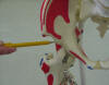
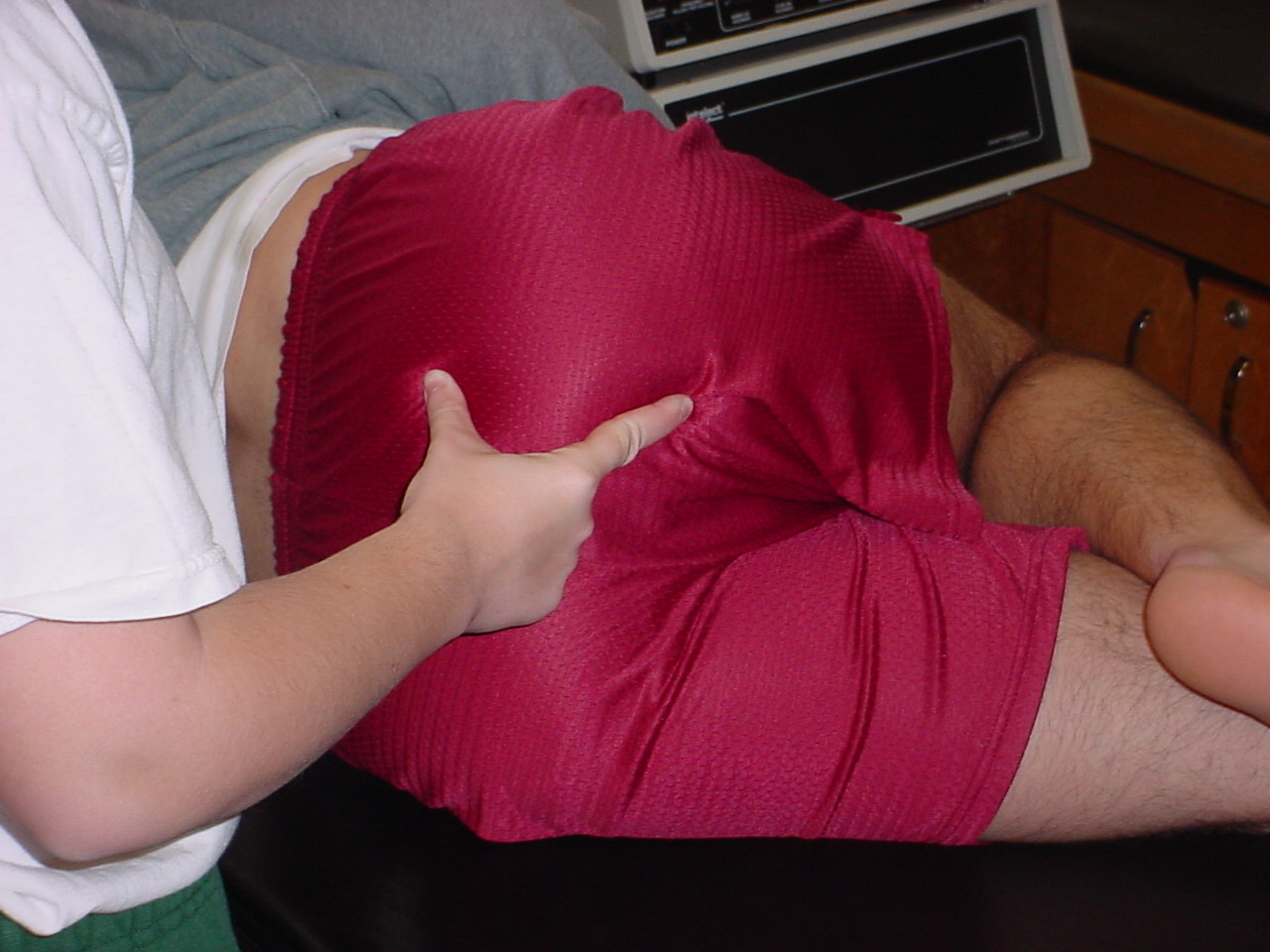
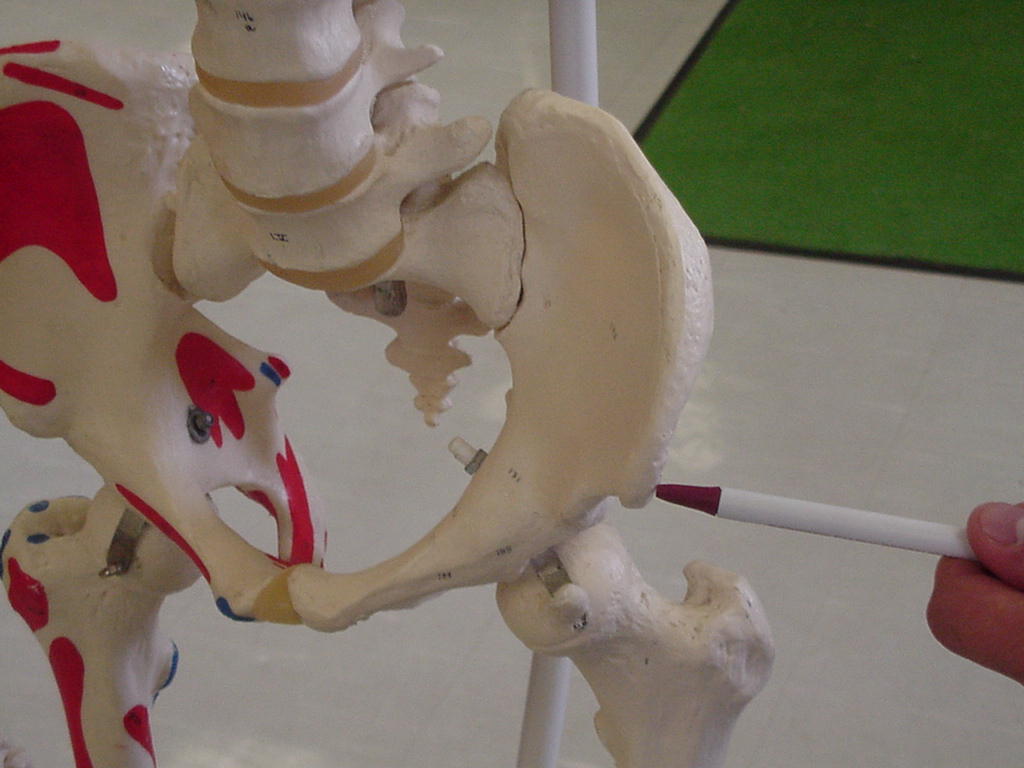
| Greater trochanter |
The
posterior edge of the greater trochanter is relatively uncovered and
easily palpable; however, the anterior and lateral portions are covered
between the tensor fascia lata and gluteus medius muscles and are less
palpable |
Patient
lying on side |
Locate
by moving fingers downward from the iliac tubercle to the greater
trochanter |
 |
 |
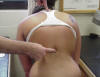
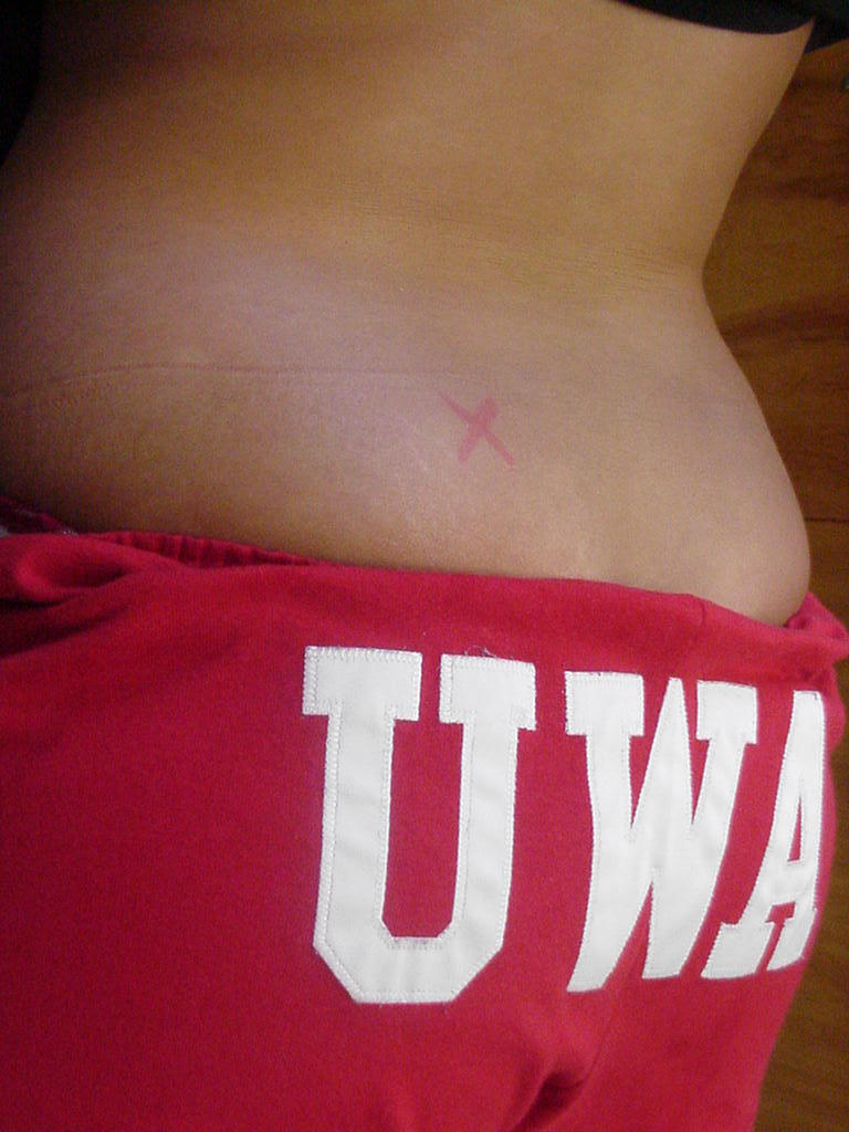
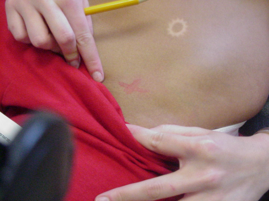
| Posterior-superior iliac spine |
Origin
of the gluteus maximus muscle |
Patient
standing or side-lying |
Lies
immediately underneath the visible dimples just above the buttocks; to
palpate, move just inferior and lateral from L5 lumbar spine and you will
feel a bony prominence this is the posterior-superior iliac spine |
 |
 |
| Ischial tuberosity |
|
Side-lying
position |
Located
in the middle of the buttocks at the approximate level of the gluteal fold |
|
 |
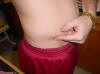
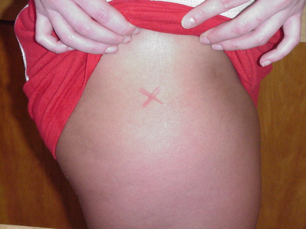
| Anterior-superior iliac spine |
Serves as a point of reference
in physical examinations |
Patient should be standing,
relaxed |
Located just distal to the
iliac tubercle |
 |
 |
| Anterior-inferior iliac spine |
Origin
for the rectus femoris muscle |
Standing
erect or supine |
Located
inferior to the anterior superioriliac spine |
 |
 |
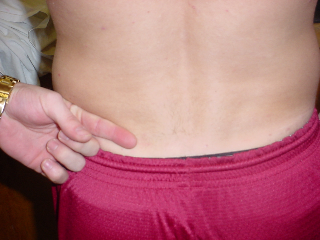
| Crest of ilium,
Iliac crest |
Origin
of gluteus medius and tensor fasciae latae |
Side
lying on unaffected side with knee flexed to 70° |
Superior
to the anterior superior iliac spine and medial to the iliac tubercle |
_small.JPG) |
 |
| Iliac tubercles |
The iliac tubercle serves as
the widest point of the iliac crest |
Patient should be standing,
relaxed |
Bony prominence located
posterior to the iliac crest |
|
|
| Posterior-inferior iliac spine |
Origin
of gluteus maximus |
Prone |
Inferior
to posterior superior iliac spine and superior to sciatic notch |
_small.JPG)
XX |

XX |
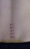
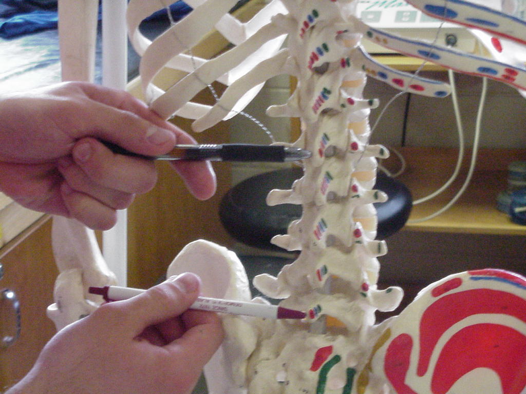
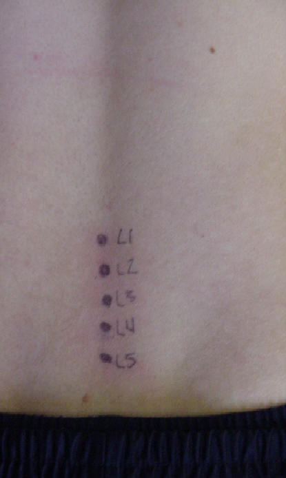
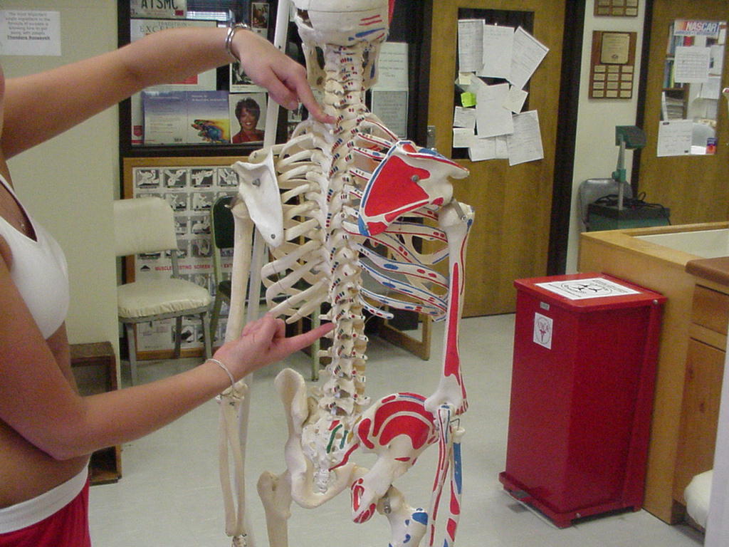
| Lumbar spine spinous process
(L1-L5) |
Serves
as an attachment for the supraspinous and the interspinous ligaments |
Patient
lying prone and relaxed |
L5
is located between the PSISs then move one finger width upward |

XX |
 |
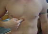
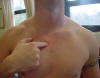
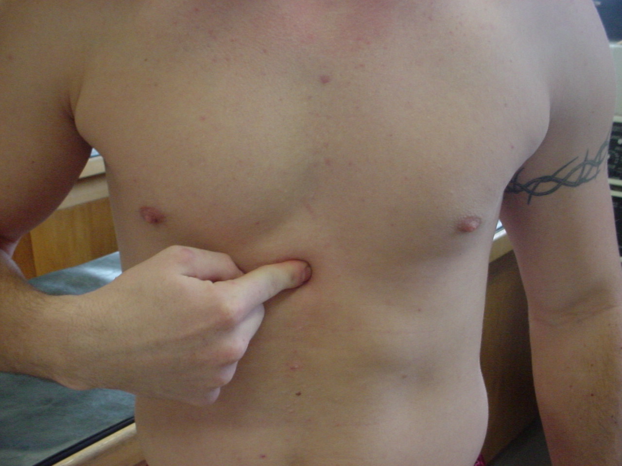
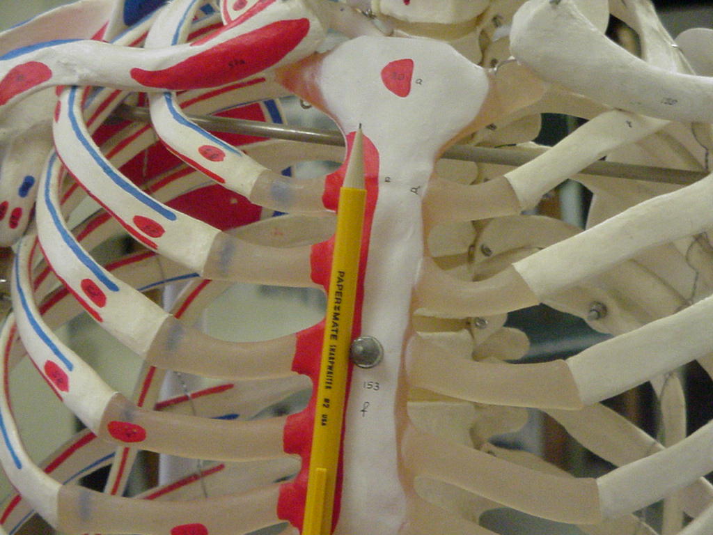
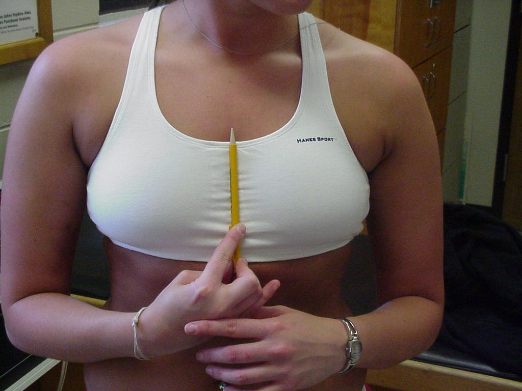
| Xiphoid process |
Attachment
site of rectus abdominis, aponeuroses of internal and external obliques,
linea alba, and diaphragm |
Standing
erect with shoulders retracted |
Inferior
portion of the sternum |
_small.JPG) |
 |
| Pubic tubercles |
Serves as an attachment for
the adductor longus muscle |
Patient should be standing,
relaxed |
Located anteriorly and
inferiorly to the anterior superior iliac spine |
 |
|
| Thoracic spine spinous process
(T1-T-12) |
The
skeletal foundation of the thorax is formed by 12 pairs of ribs that
insert on the spine |
Patient
can be prone or standing |
T-1-T-12
are respectively located directly inferior to C-7 |
 |
 |
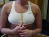
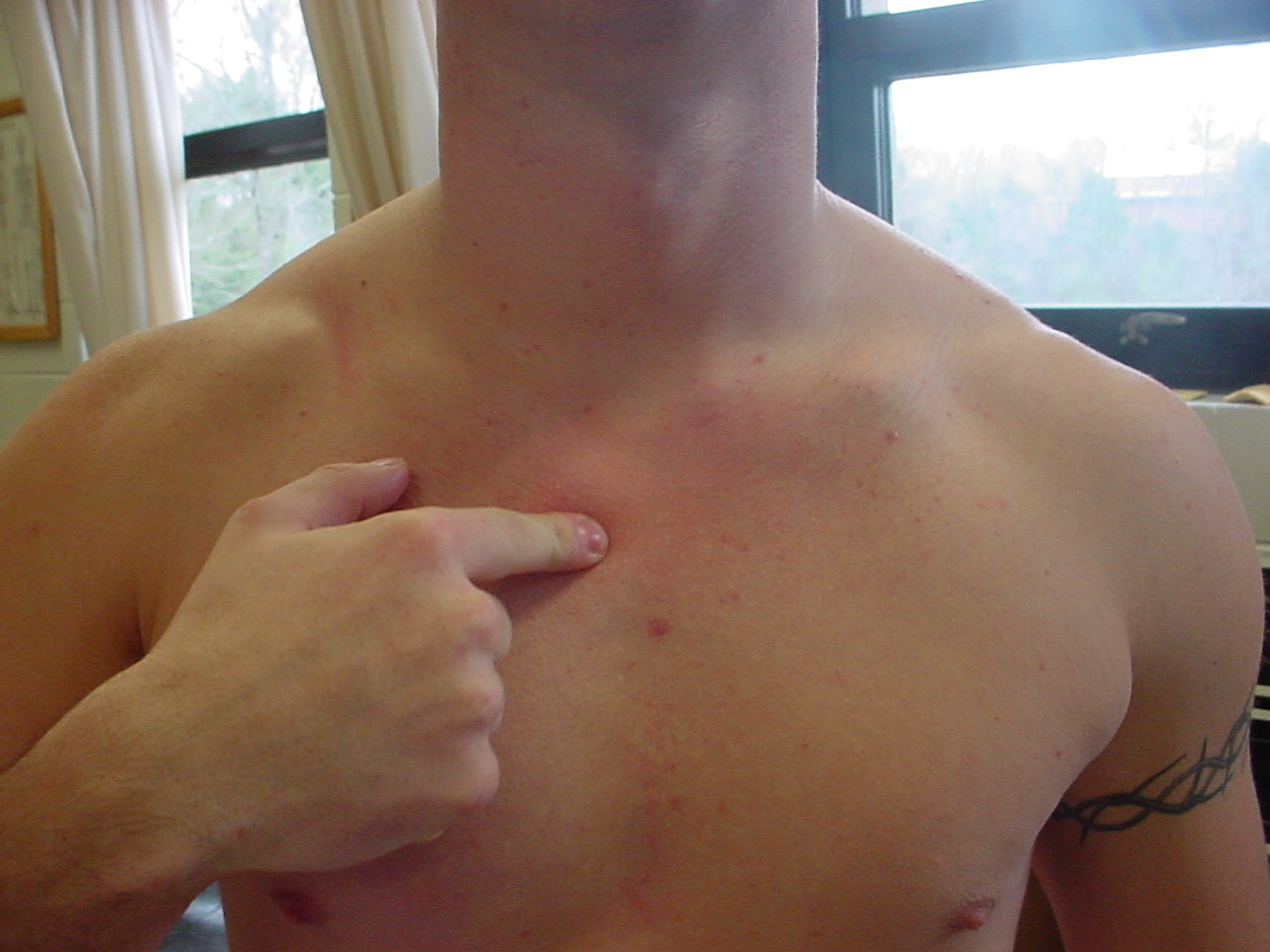
| Sternal body |
Serves
as an attachment for the costal cartilages of the ribs 2-10 |
Patient
can be standing, seated, or supine |
Located
inferiorly to the sternoclavicular joint and manbrium, and superior to the
xiphoid process |
 |
 |
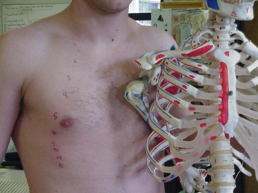
| Manubrium
or Manubrium of Sternum |
Attachment
site of clavicle to sternum (anterior sternoclavicular ligament) |
Supine |
Most
superior portion of sternum |
_small.JPG) |
 |
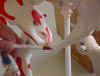
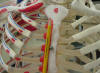
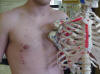
| Ribs 1-12 |
Ribs 1-7 join the sternum
directly by their costal cartilages. The cartilages of the false ribs join
the cartilage of the seventh rib. The ribs support the bones of the body
and protect internal organs |
Patient should be lying in
the supine position and relaxed. |
The ribs are located lateral
to the ribs |
 |
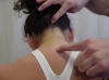
| Cervical spine spinous process
(C2-C7) |
C-2
is also called the "axis", C-7 near the tip of its spine serve
as an attachment site for a number of the upper back and shoulder muscles |
Patient
standing, seated, or supine |
C-2 is
the pivot on which the head rotates, inferior to the atlas or C-1; C-7 is
located by the prominent bump on the posterior surface of the neck or
cervical region |
 |
 |
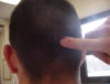
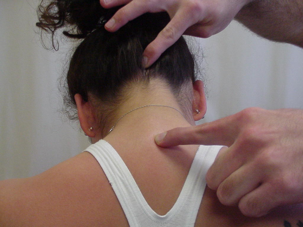
| Occipital protuberance |
|
|
|
_small.JPG) |
 |


.JPG)

.JPG)
.JPG)


.JPG)

.JPG)
