|
Bony Landmark
(include alternative name if applicable)
|
Related Information
such as purpose, function,
attachment of ligaments, tendon, soft tissues involved
|
Preferred Body &
Joint Position
best for palpation
|
Anatomical
Description of Location
relative to other structures
|
Skeleton Picture or Video
|
Model Picture or Video
|
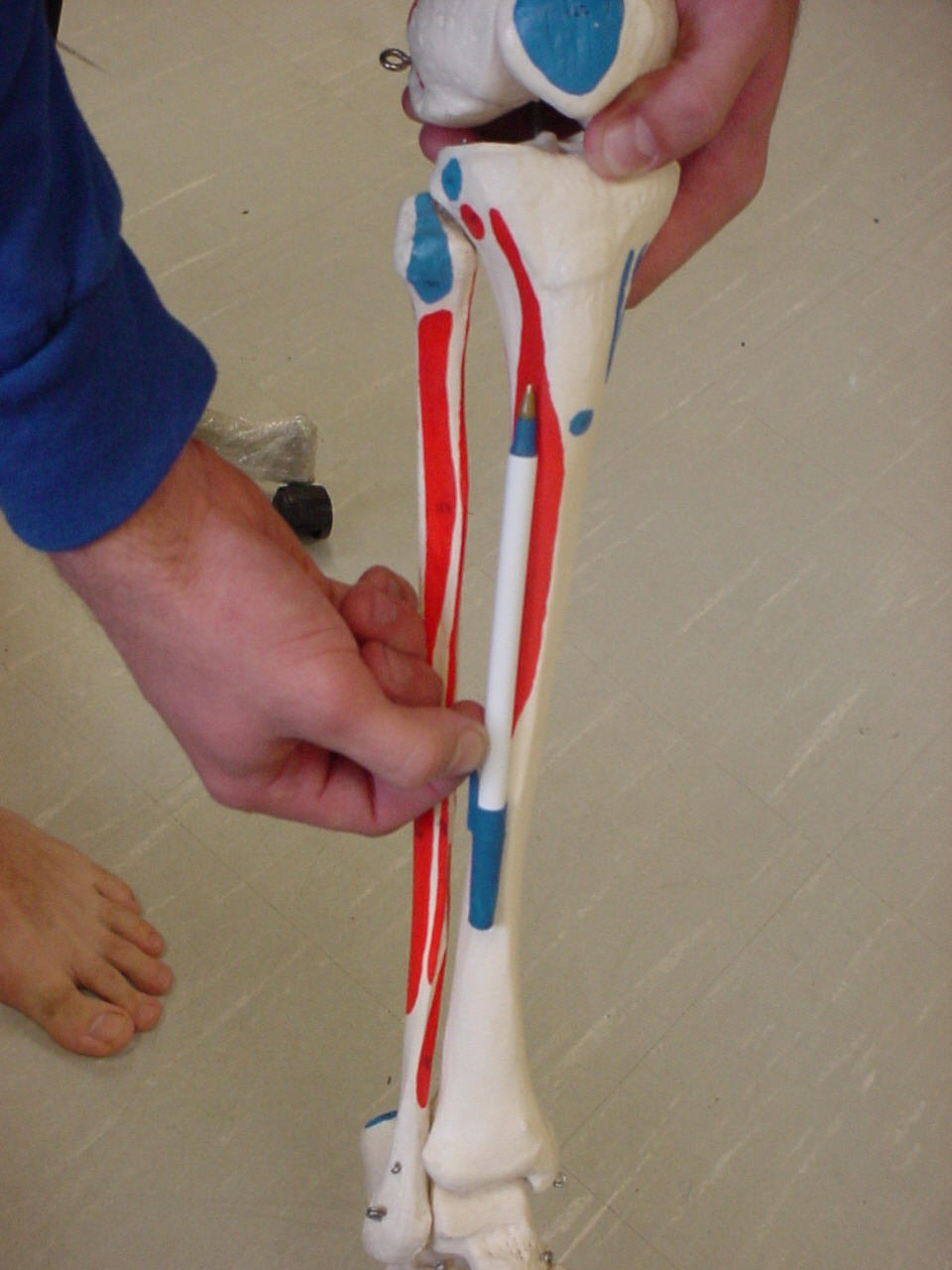
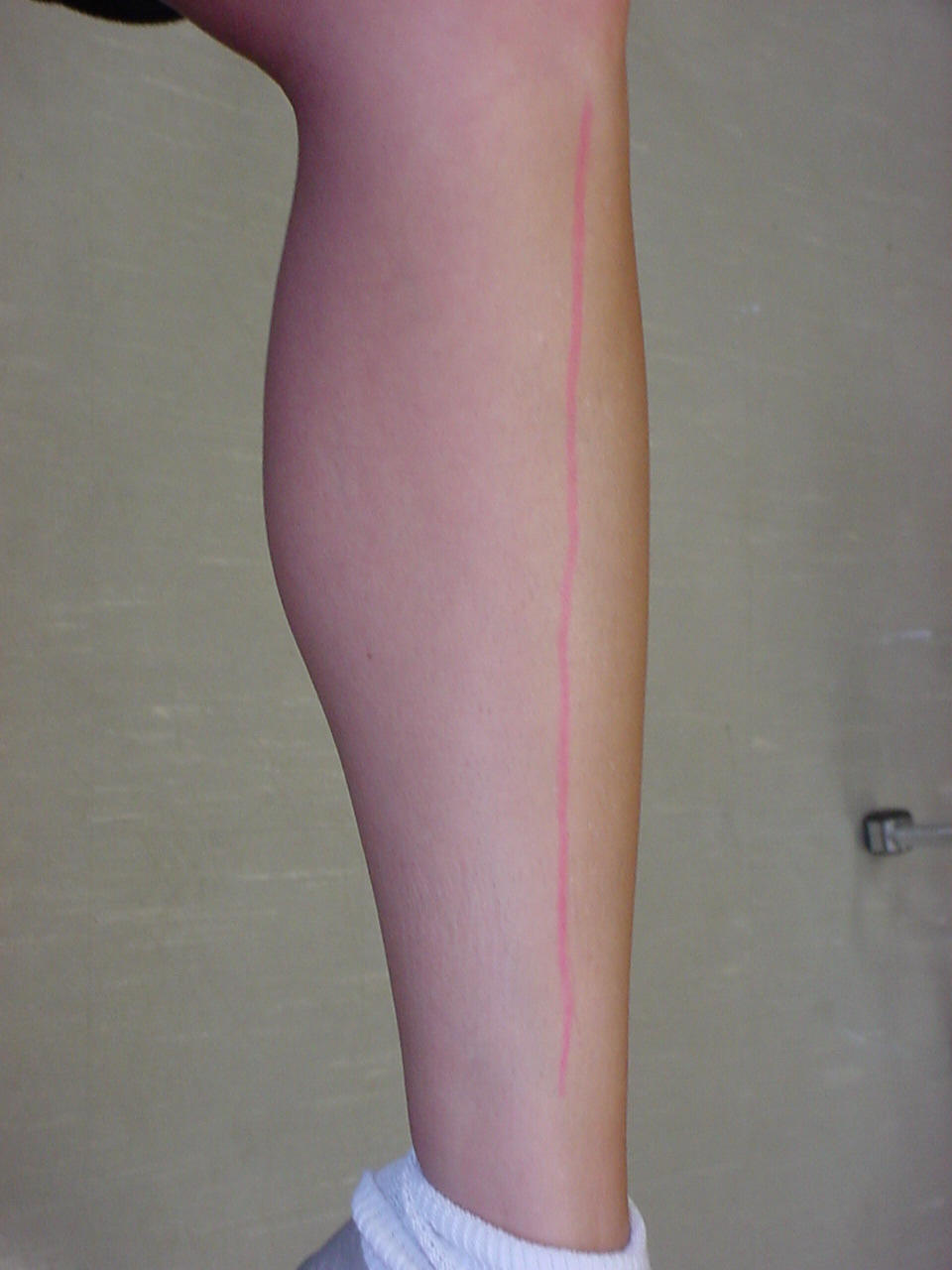
| Tibial Spine |
Origin of the
tibialis anterior muscle |
Patient seated |
Located on the anterior portion
of the lower leg; It can be palpated from just above the ankle to just
below the knee; The tibial spine runs along the midline of the anterior
portion of the leg |
 |
 |
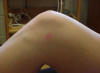
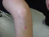
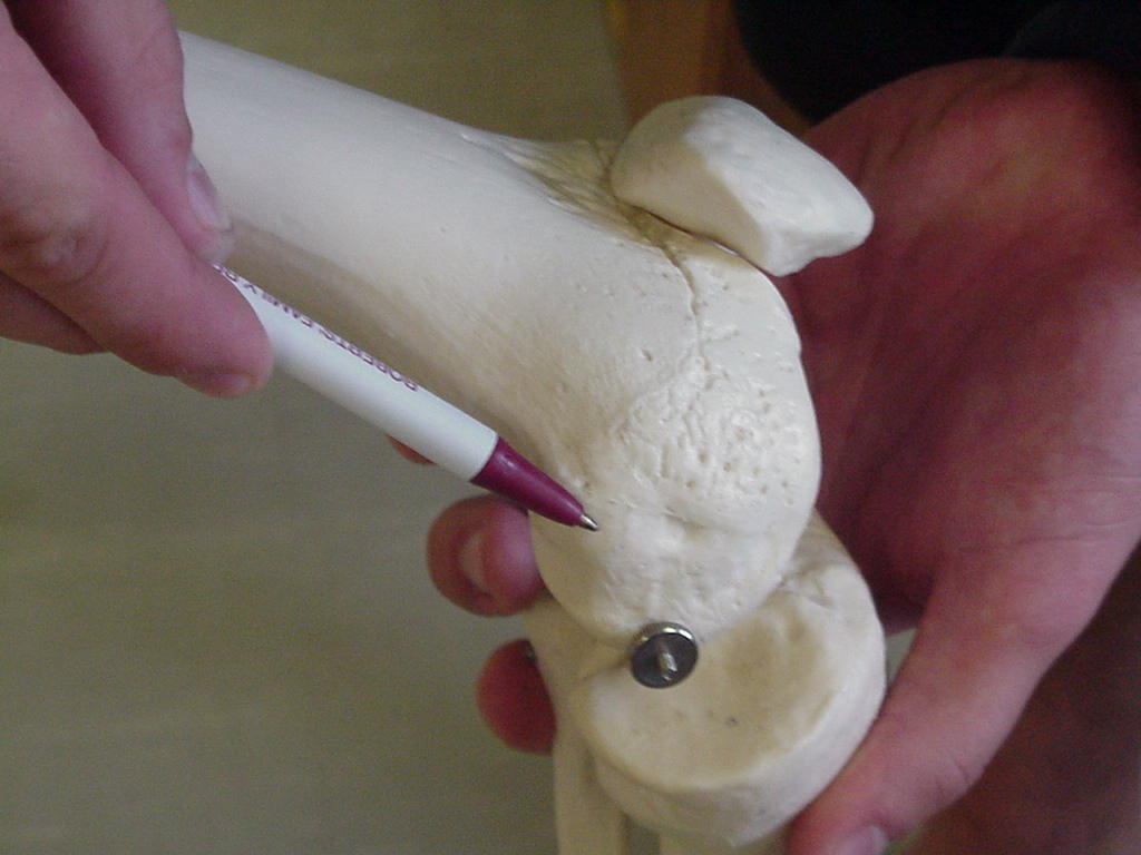
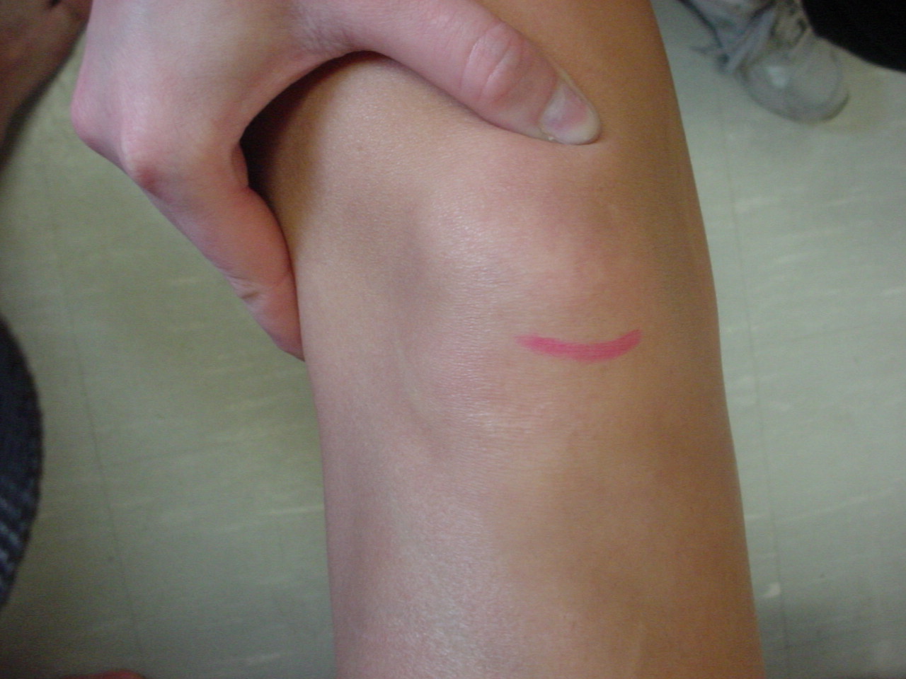
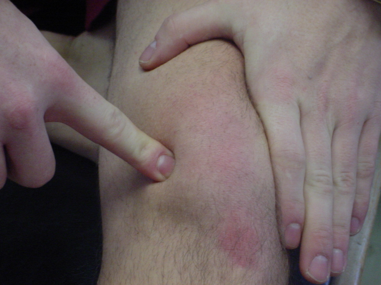
| Lateral tibial plateau |
Site of lateral
meniscus |
Short-sitting position |
Superior lateral
portion of the tibia |
|
|
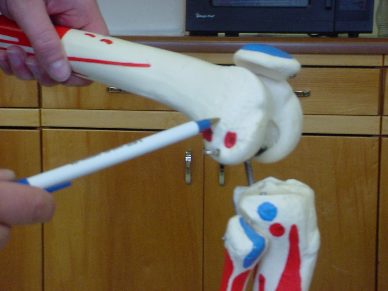
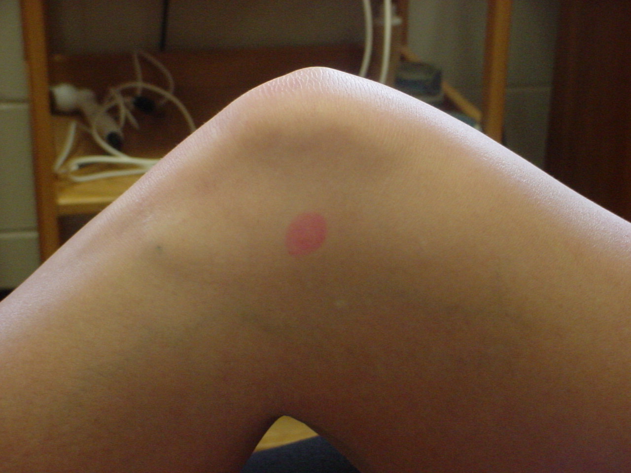
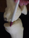
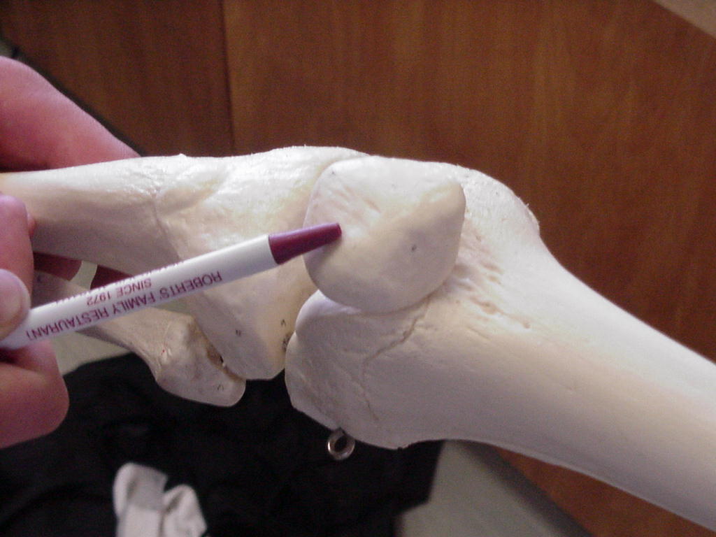
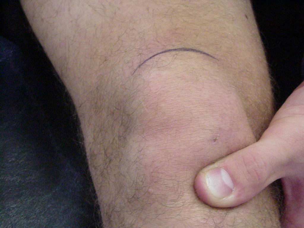
| Fibula head |
Attachment site of
the distal end of the the fibular collateral ligament; the fibular
collateral ligaments attaches the fibula to the femur |
Patient in supine
position with the knee flexed to 90° |
Approximately 1cm
inferior and posterolateral to the lateral tibial plateau |
|
|
| Medial femoral condyle |
Serves as an attachment for the medial
collateral ligament |
Patient sitting on the edge of the table with
knee flexed 30 degrees and with the patient being relaxed |
The medial femoral condyle can be palpated
proximally as far as the superior pole of the patella and distally where
the femur and the tibia meet |
 |
|
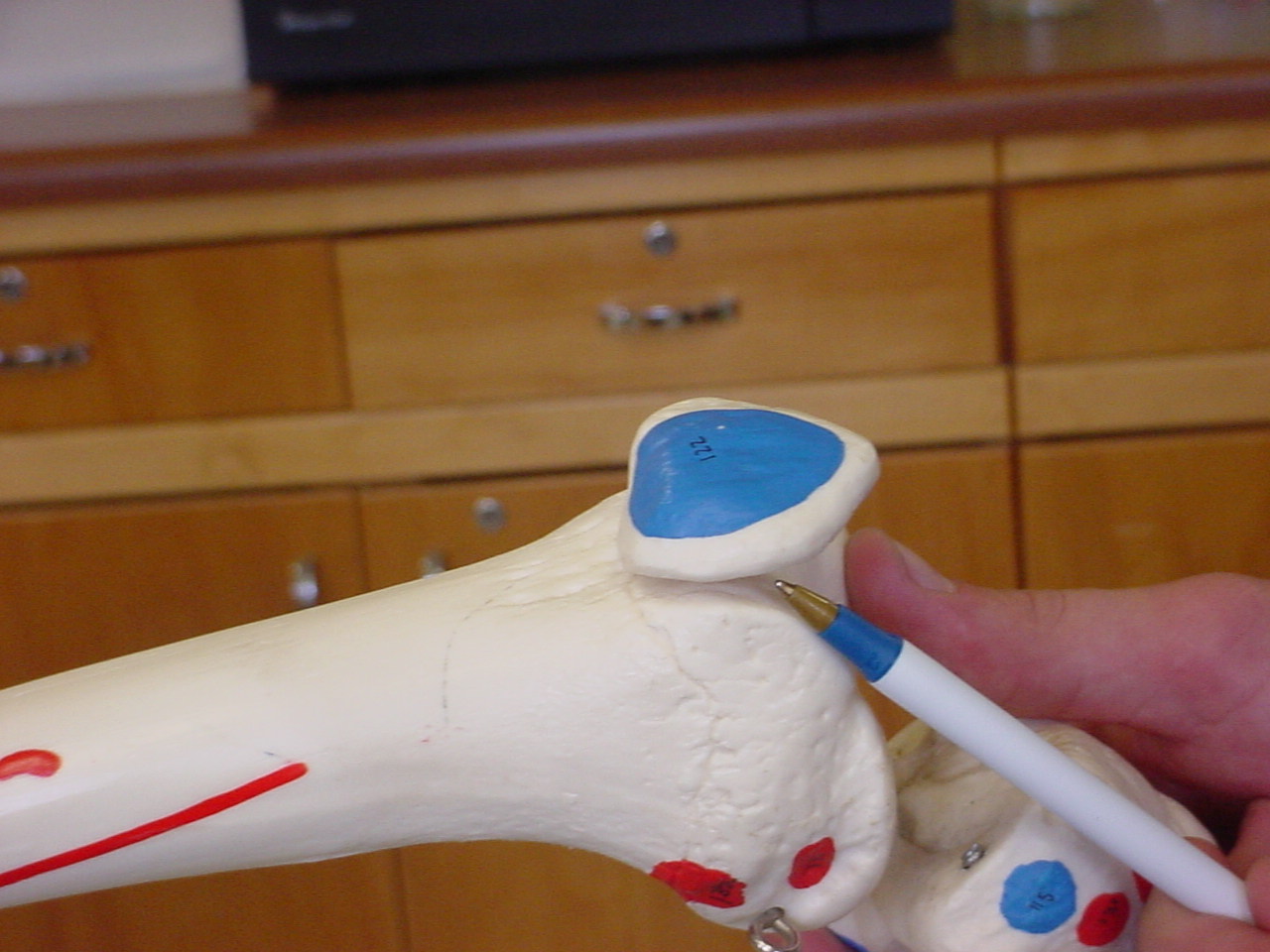
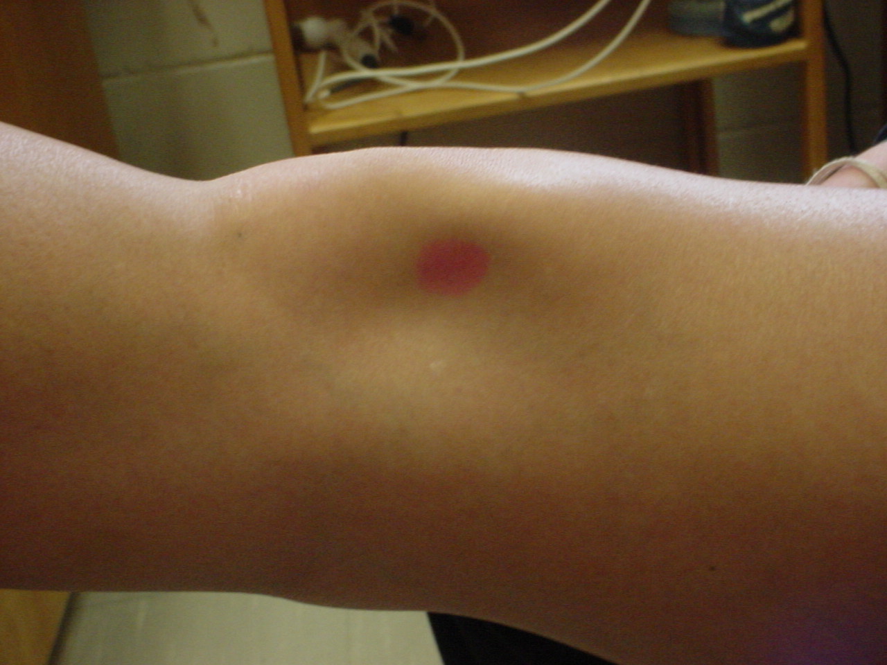
| Patella superior pole |
Attaches to the quadriceps to form the quadriceps tendon |
Patient sitting on the edge of the table with knee flexed
approximately 30 degrees |
Just superior to the patella tendon |
 |
 |
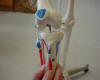
| Patella |
It functions in lever actions associated with
lower limb movements. The patella serves as an attachment for the
quadriceps muscle group |
Patient needs to be relaxed in the supine
position with leg in extension |
The patella is located in a tendon that passes
anteriorly over the knee |
 |
 |
| Patella inferior pole |
Patella tendon
crosses patella or some say it is were the patella tendon originates |
Patient seated or
standing, with the knee flexed or straight |
Located at the
extreme inferior aspect of the patella
|
 |
 |
| Patella lateral facet |
Helps to hold the
quadriceps tendon away from the axis of movement, it also functions as a
guide for the quadriceps tendon of the knee, and provides protection for
the cartilage of the femoral condyles |
Model sitting with
knees off the end of a table |
It is fixed in the
trochlear groove in flexion and mobile in extension |
 |
 |
| Patella medial facet |
Attachment of
patella ligament and medial patella retinaculum |
Short-sitting
position |
Articulates with
the medial condyle of the femur |
_small.JPG) |
 |
| Trochlear groove |
Location of
patellar tracking |
Supine |
Located beneath the
patella |
_small.JPG) |
|
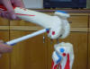
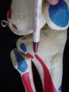
| Lateral femoral condyle |
Origin of the
popliteal muscle; Much of it is covered by the patella causing less
palpable surface area |
Flex the knee past
90 degrees; the patient can be prone or seated |
Find the soft
tissue depression lateral to the infrapatellar tendon; move superior and
laterally onto the sharp edge of the lateral femoral condyle |
 |
 |
| Medial tibial plateau |
Attachment for the medial meniscus |
Patient sitting on edge of table with knee
flexed approximately 30 degrees |
Located in the soft tissue depression on the
anterior portion of the knee, just medial of the patella tendon |
 |
 |
| Adductor tubercle |
Attachment of
adductor magnus tendon |
Short-sitting
position |
Posterior medial
portion of the medial femoral condyle |
|
|
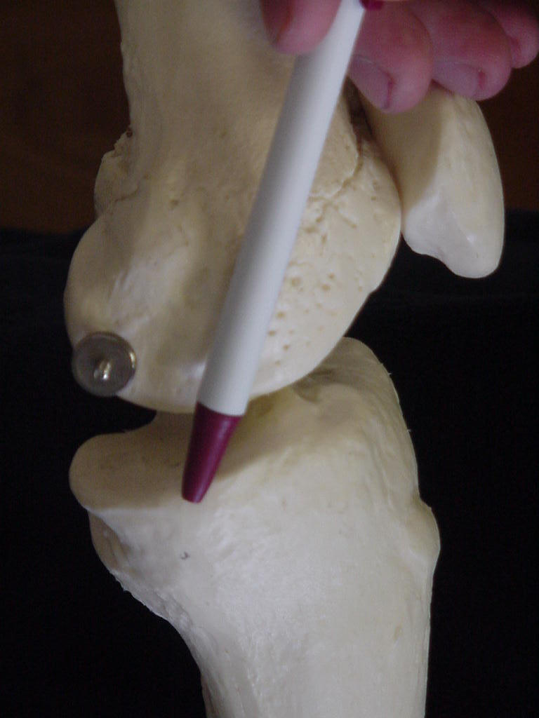
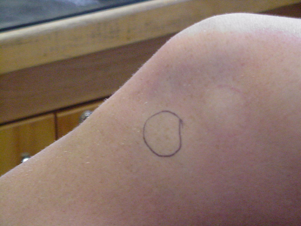
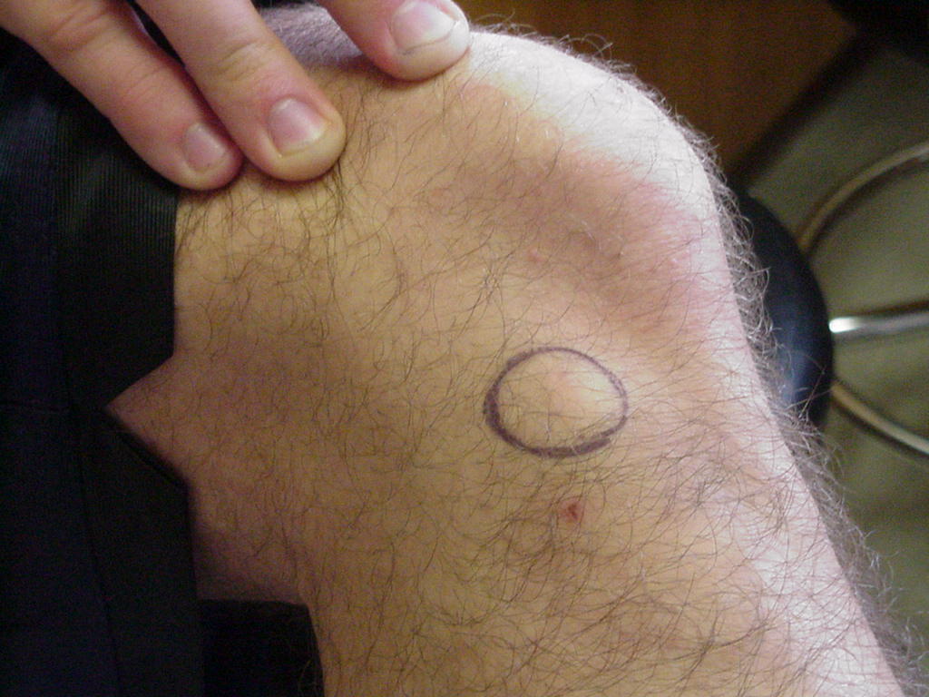
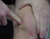
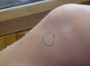
| Tibial tubercle |
Attachment of the
patellar tendon, the pes anserine inserts here, and the also a bursa |
Patient needs to be
sitting or supine |
Located at the
upper end of the anterior border of the shaft of the tibia, and is the
apex of the triangular area on the front of the bone where the anterior
surfaces of the two condyles become continuous, Follow the infrapatellar
tendon distally to where it inserts into the tibial tubercle |
 |
 |
| Gerdys tubercle |
Insertion for the Iliotibial Tract |
Patient sitting on the edge of table with knee
flexed approximately 30 degrees and relaxed |
Located immediately below the lateral tibial
plateau |
 |
 |


.JPG)
.JPG)