|
Bony Landmark
(include alternative name if applicable)
|
Related Information
such as purpose, function,
attachment of ligaments, tendon, soft tissues involved
|
Preferred Body &
Joint Position
best for palpation
|
Anatomical
Description of Location
relative to other structures
|
Skeleton Picture or Video
|
Model Picture or Video
|
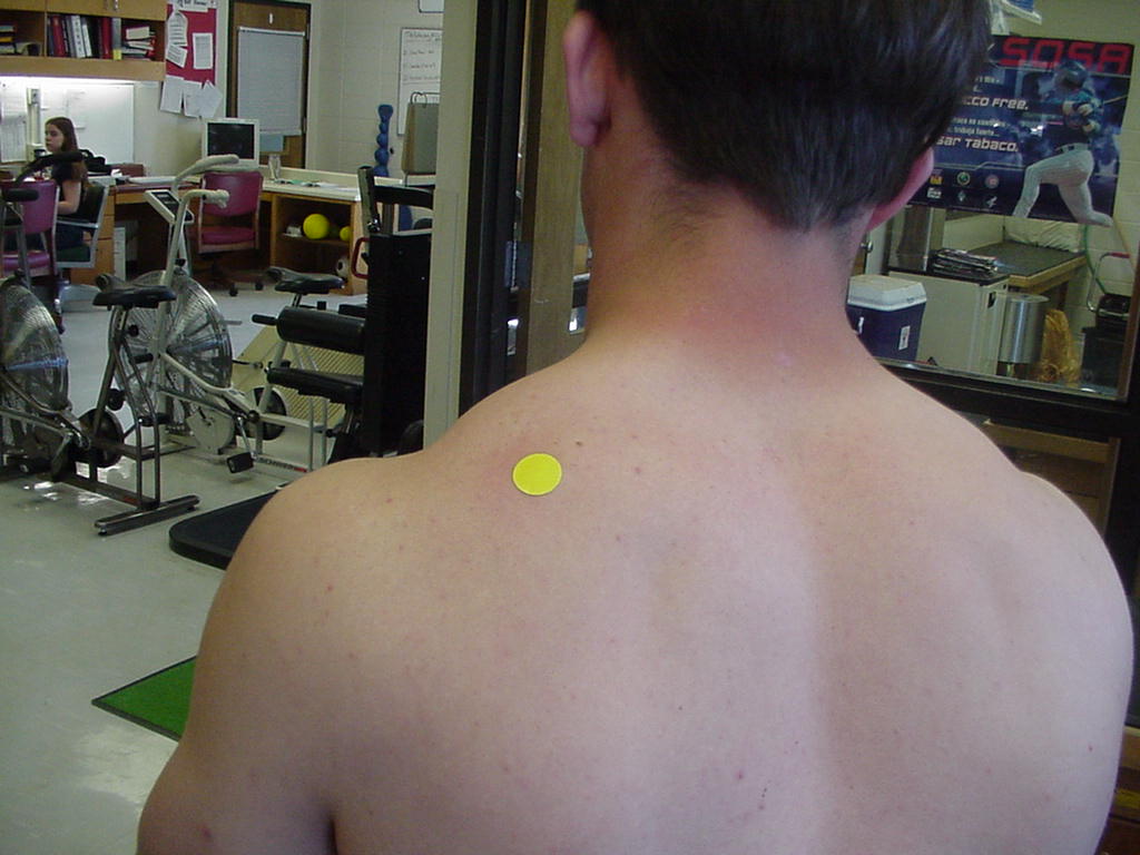
| Scapula medial (vertebral) border |
Attaches to ribs 2-7 |
Patient standing, arms by side, relaxed |
Located 2" from the spinous processes |
|
|
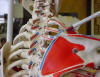
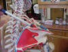
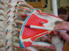
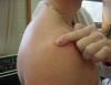
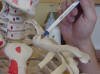
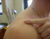
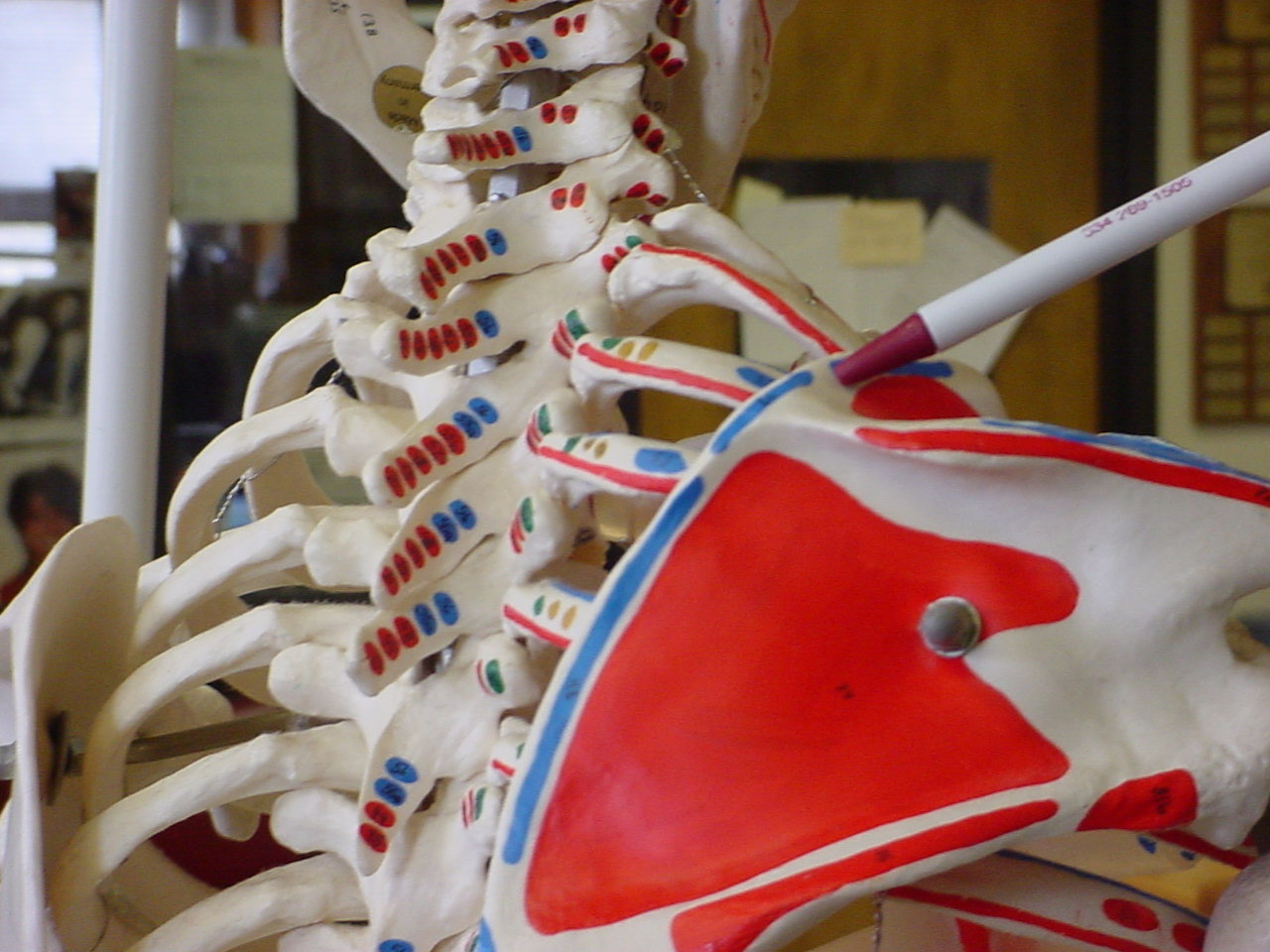
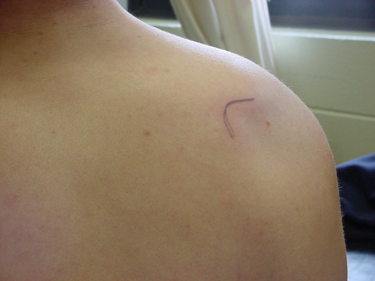
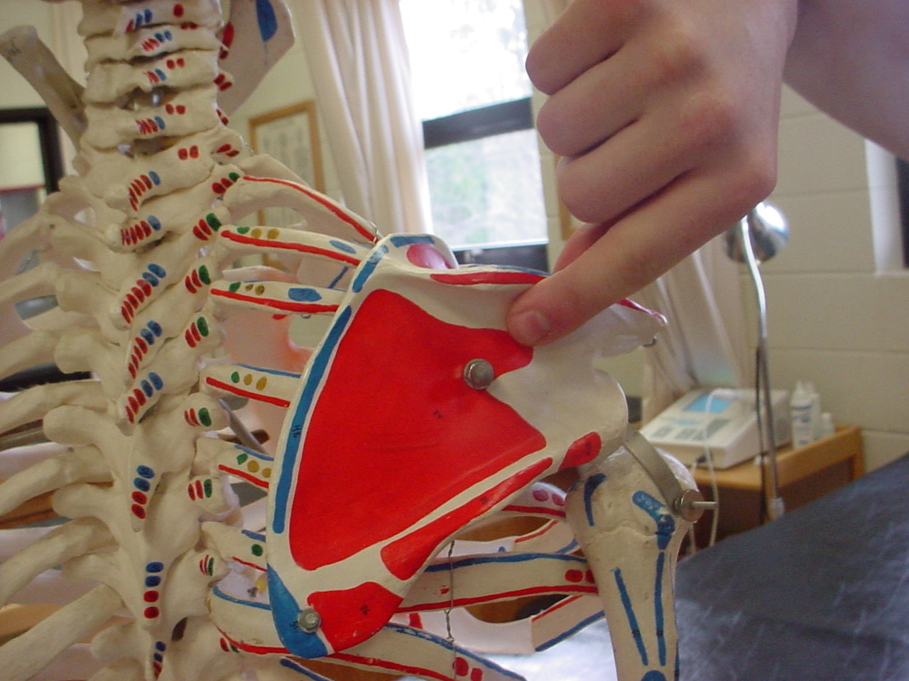
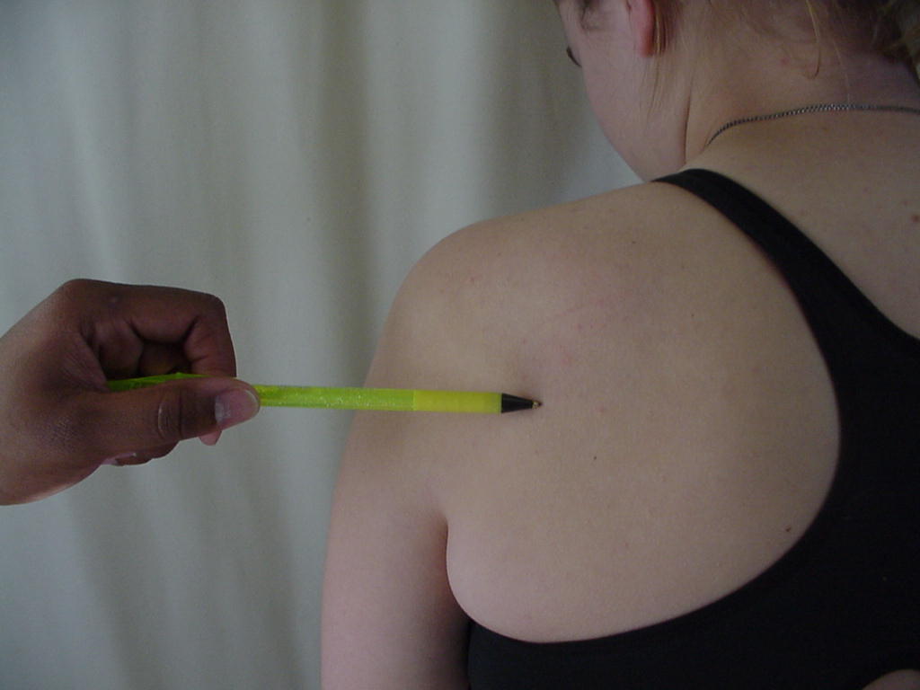
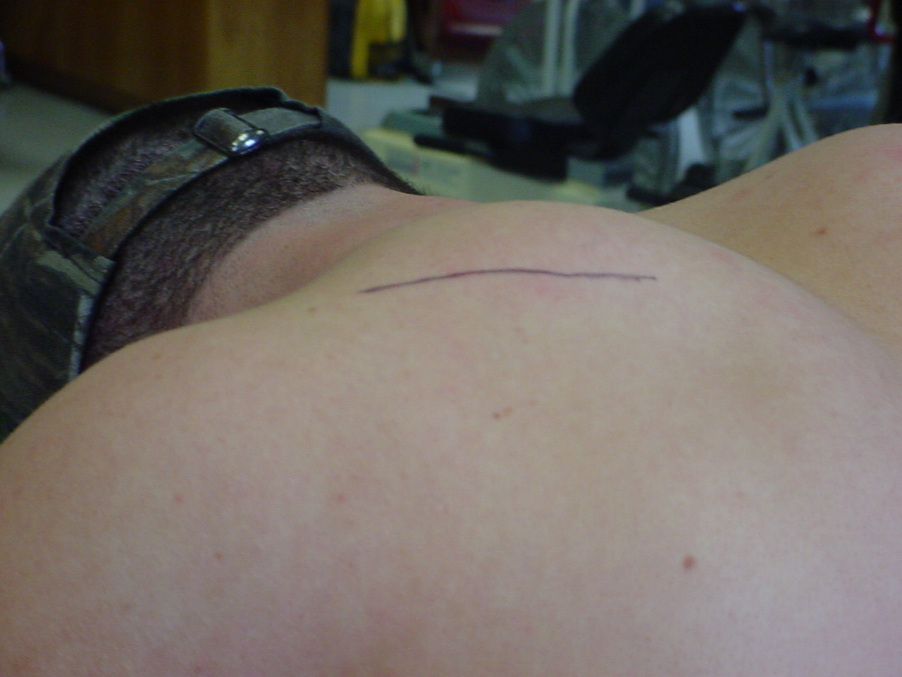
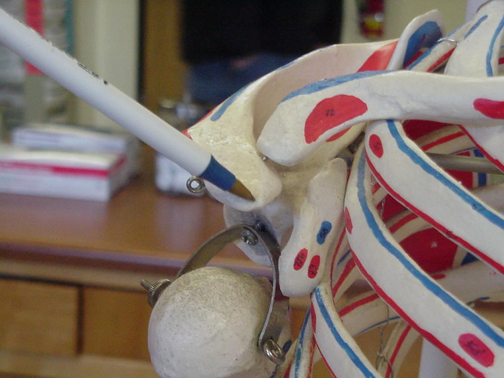
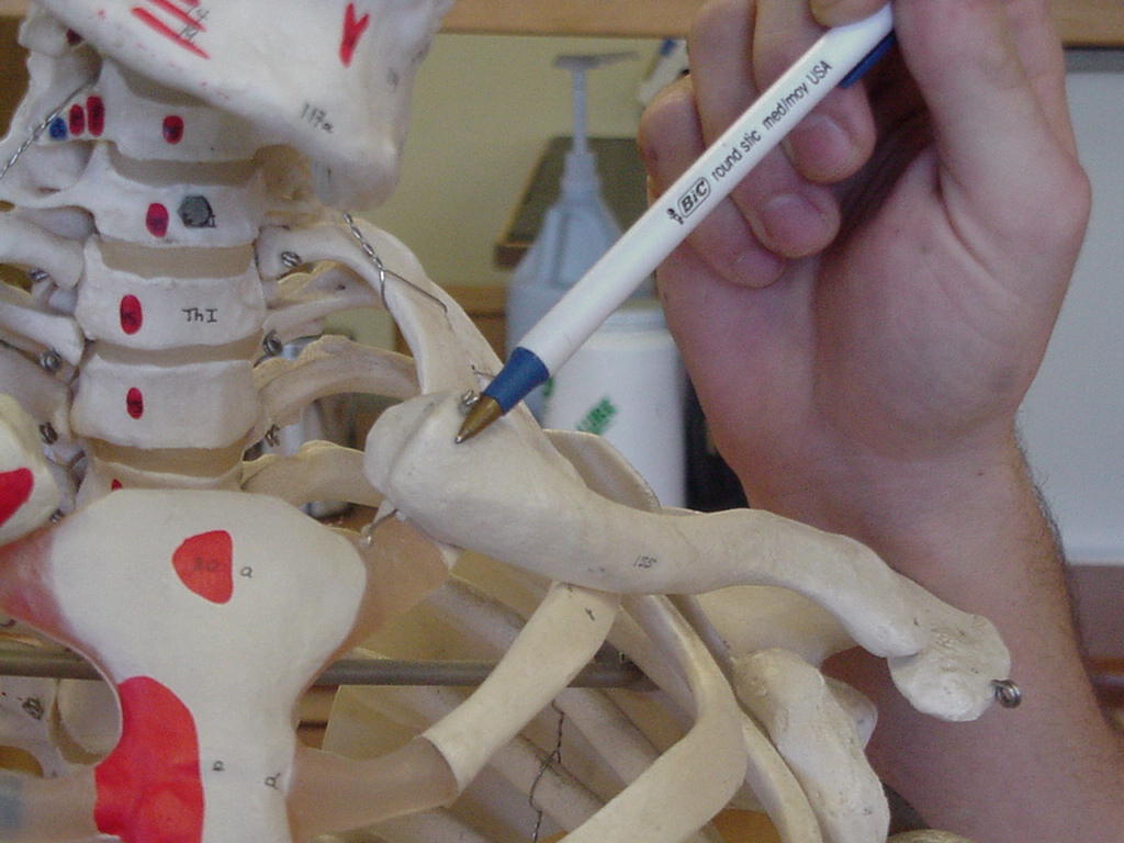
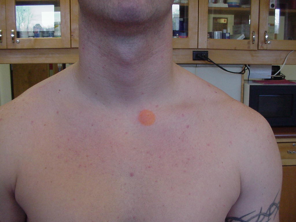
| Scapula lateral (axillary) border |
Origin of the
triceps brachii long head, teres minor, and teres major muscles |
Position the
patient with the elbow flexed about 90 degrees and behind the back; the
patient may be seated or standing |
From the most
inferior point of the scapula move superior and lateral along the scapula,
this makes up the lateral border |
BorderSkeleton_small.JPG) |
BorderBody_small.JPG) |
| Scapula inferior angle |
Origin of teres
major |
Prone |
Inferior portion of
scapula |
_small.JPG) |
 |
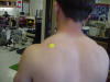
| Scapula superior angle |
Insertion of serratus anterior muscle, levator
scapula muscle, and attachment for supraspinatus muscle |
Patient standing, arms by side, relaxed |
Located just lateral to the 2nd rib and
superior to the spine of the scapula |
XX |

XX |
| Supraspinatus fossa |
Fossa in which the
supraspinatus muscle is located |
Patient sitting or standing with back to the
examiner |
Located just superior to the spine of the
scapula |
 |
 |
| Infraspinatus fossa |
Formed by the space
between the infraspinatus muscle and the spine of the clavicle |
Prone |
Inferior to the
spine of the scapula |
 |
 |
| Spine of scapula |
The spine of the scapula serves as the
attachment for the deltoid and the trapezius muscles |
The patient should be standing and relaxed |
The spine of the scapula is a sharp
subcutaneous ridge running diagonally across the posterior surface of the
shoulder blade |
 |
 |
| Coracoid process |
Origin for the
Corocobrachialis muscle |
Standing erect or
seated |
Press laterally and
posteriorly under the anterior edge of the clavicle about one inch from
the lateral end of the clavicle |
 |
 |
| Acromion process |
Articulates with
the clavicle (forms the AC joint); coracoacromial ligament attaches here; |
Sitting or standing |
Distal to the
lateral 1/3 of the clavicle; "summit of the shoulder;" |
_small.JPG) |
 |
| Clavicle |
Serves as an attachment for the pectoralis
major, deltoid, and trapezius muscles |
Patient sitting in a chair with the examiner
sitting behind them |
Located lateral from the sternoclavicular joint |
|
|
| Clavicle sternal end |
Fits into the
manbrium of the sternum to form the sternoclavicular joint |
Patient can be
seated, standing, or supine |
Located by
following the clavicle medially until you get to the nodule that sticks
out (which is the insertion for the sternocleidomastoid muscle); It is
located just before the trachea. |
 |
 |
| Clavicle acromial end,
AC joint or acromioclavicular joint |
Supported by the
coracoclavicular ligaments (trapezoid and conoid); superior and inferior
AC ligaments also provide stability |
Short-sitting or
standing |
Where the distal
end of the clavicle articulates with the acromion |
_small.JPG) |
 |
BorderSkeleton.JPG)
BorderBody.JPG)
.JPG)


.JPG)
.JPG)
