|
Bony Landmark
(include alternative name if applicable)
|
Related Information
such as purpose, function,
attachment of ligaments, tendon, soft tissues involved
|
Preferred Body & Joint
Position
best for palpation
|
Anatomical Description of Location
relative to other structures
|
Skeleton Picture or Video |
Model Picture or Video
|
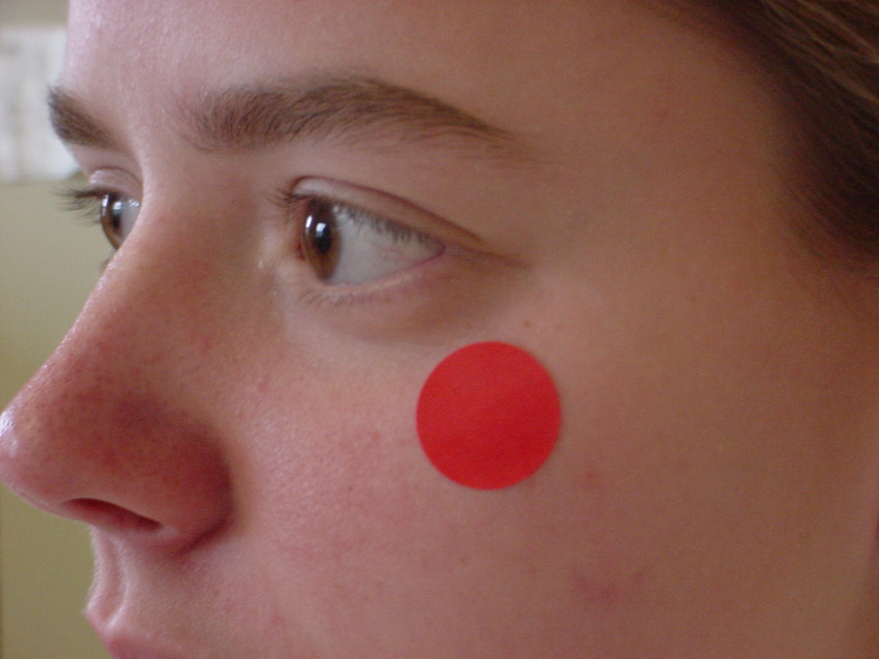
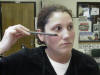
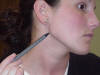
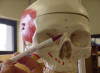
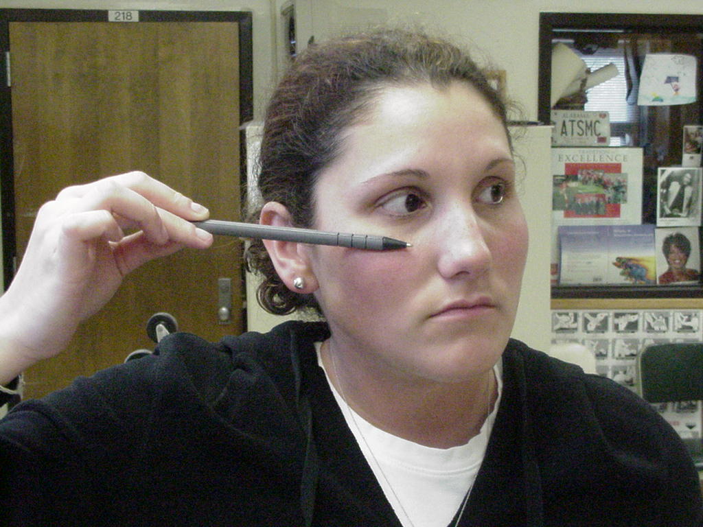
| Lateral
Orbital Margins- outer margin of orbit formed by the malar or zygomatic
bone |
|
Patient needs to be
seated, standing, or supine |
Immediately lateral
to the eye orbital socket, outer origin of the eye orbit formed by the
malar or zygomatic bone |
 |
 |
| Lower
border of Mandible- bony ridge of the jawbone |
Bony ridge of the
jaw bone |
Patient should be
facing the examiner either standing or sitting |
Located laterally
and inferior on the mandible |
 |
 |
| Angle
of Mandible- where mandible changes direction |
Just below is a
common fracture site for the jaw bone |
Patient standing,
seated, or supine |
Located at the
bottom of the jaw and forms almost a 90 degree angle |
 |
 |
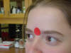
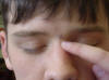
| Supraorbital
Margin- Frontal bone- ridge over each eye orbit |
Forms outer,
superior surface of eye orbit |
Eyebrows raised |
Superior part of
the eye orbit, inferior part of the frontal bone |
_small.JPG) |
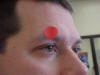 |
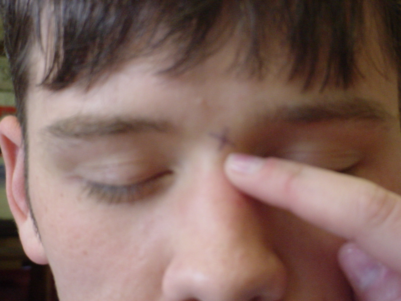
| Glabella |
Articulates with
the nasion |
Supine |
Prominent ridge just above nasion |
_small.JPG) |
 |
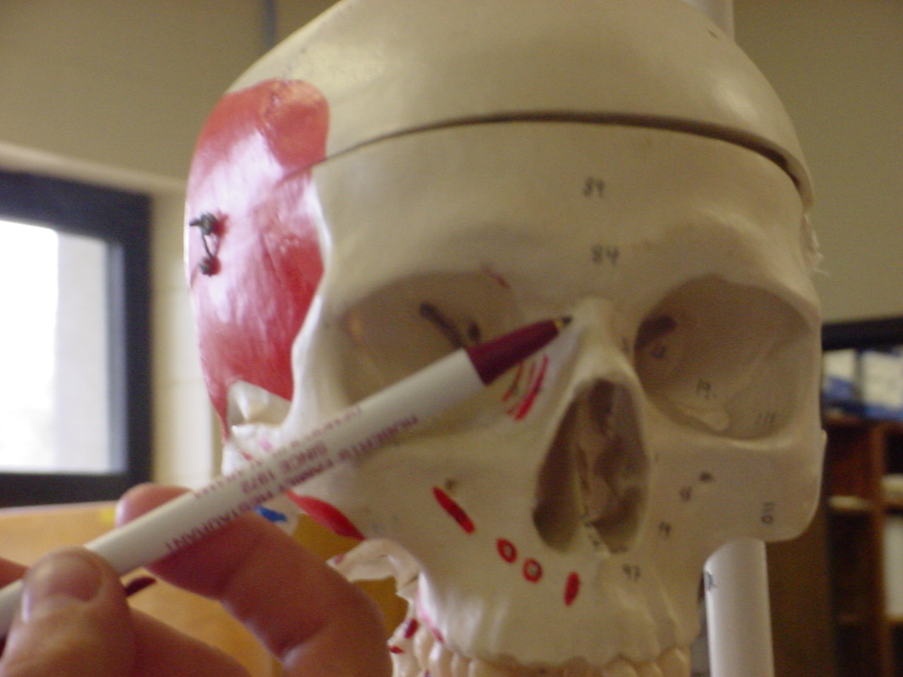
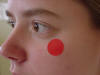
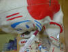
| Zygomatic
arch- prominent cheek bone |
Superficial
temporal artery crosses the posterior extremity of the zygomatic arch; a
pulse is palpable here |
Seated or standing |
Inferior and
lateral to the orbit of the eye |
_small.JPG) |
 |
| Nasion-
depression just above the nose between the supraorbital margins |
The nasion attaches to the nasal bones where
they are surrounded by the frontal bone to form the roof of the nose |
The patient sitting in a chair with the
examiner sitting behind the patient |
Located inferior to the glabella and superior
to the nose |
 |
 |
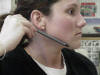
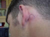
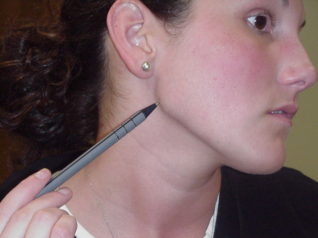
| Mastoid
processes |
Injuries to this bony prominence are
indications for Battle's Sign |
Patient lying in a supine position and relaxed |
Located posterior to the ear |
 |
 |


.JPG)

.JPG)

.JPG)